. 2007 Jul 11;27(28):7447-58.
doi: 10.1523/JNEUROSCI.4266-06.2007.

Induction of calcium influx through TRPC5 channels by cross-linking of GM1 ganglioside associated with alpha5beta1 integrin initiates neurite outgrowth

Affiliations

Induction of calcium influx through TRPC5 channels by cross-linking of GM1 ganglioside associated with alpha5beta1 integrin initiates neurite outgrowth

Gusheng Wu et al. J Neurosci. .

Abstract

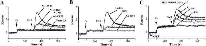
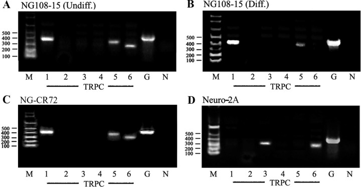
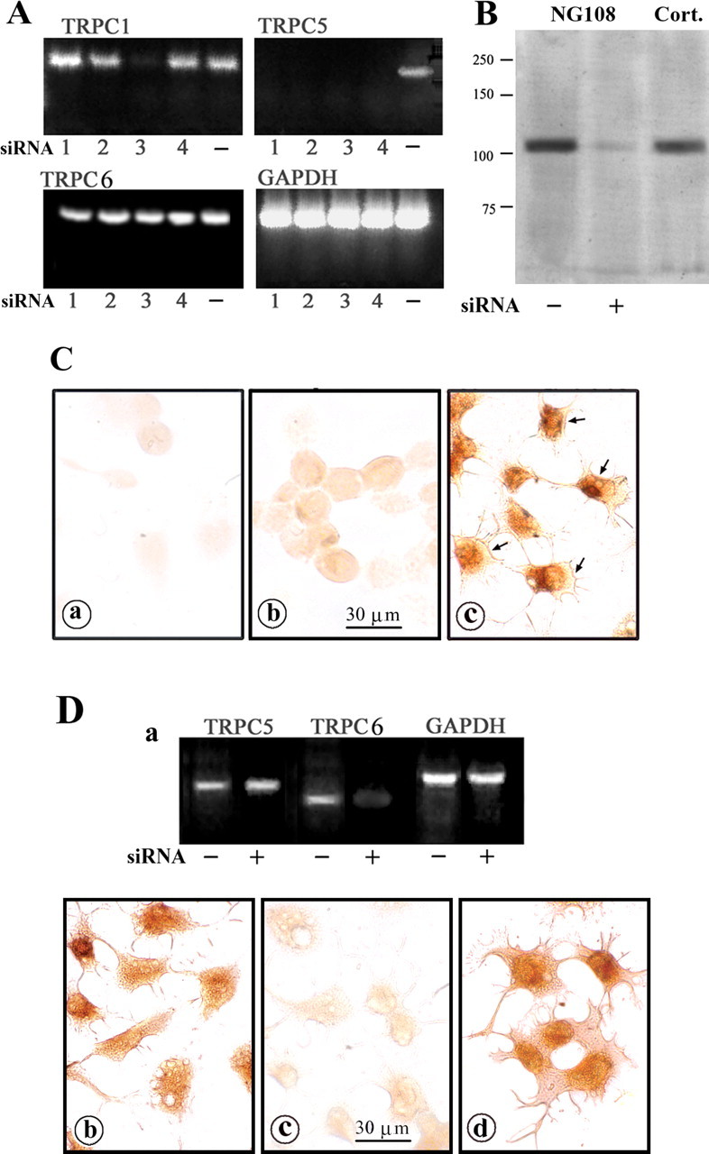
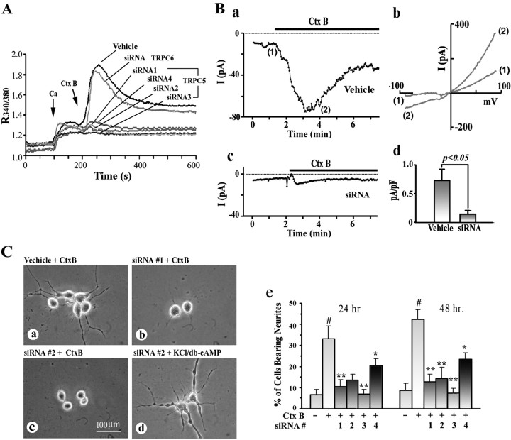
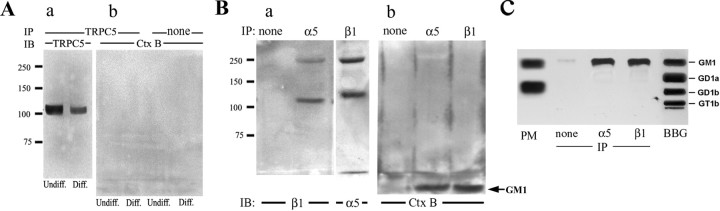
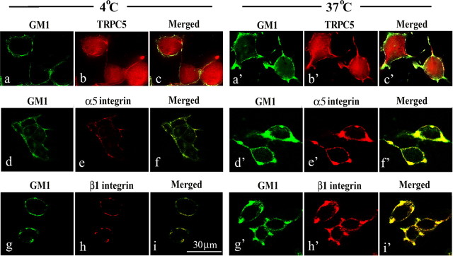
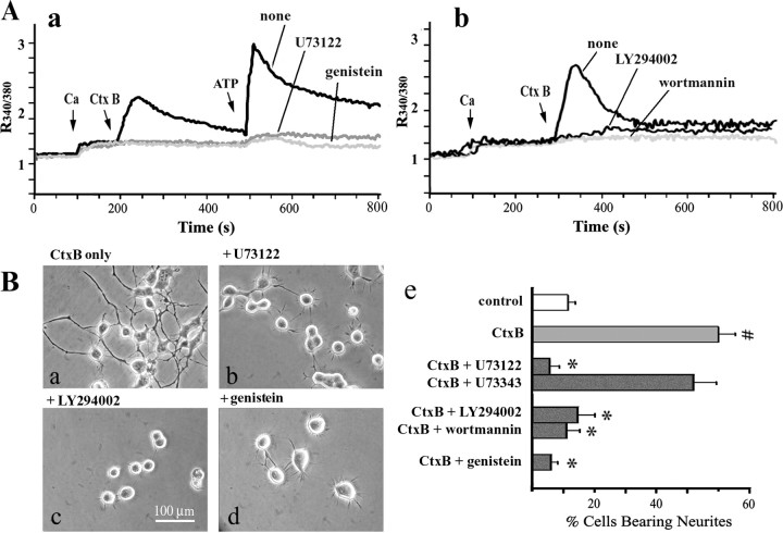
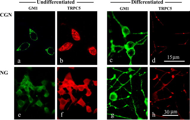
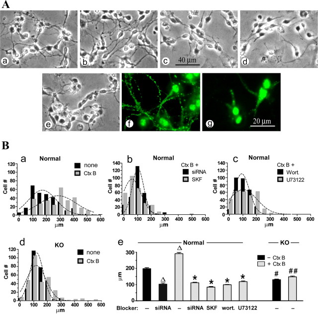
Previous studies demonstrated that cross-linking of GM1 ganglioside with multivalent ligands, such as B subunit of cholera toxin (CtxB), induced Ca2+ influx through an unidentified, voltage-independent channel in several cell types. Application of CtxB to undifferentiated NG108-15 cells resulted in outgrowth of axon-like neurites in a Ca2+ influx-dependent manner. In this study, we demonstrate that CtxB-induced Ca2+ influx is mediated by TRPC5 channels, naturally expressed in these cells and primary neurons. Both Ca2+ influx and neurite induction were blocked by TRPC5 small interfering RNA (siRNA). Pretreatment of NG108-15 cells with neuraminidase increased cell-surface GM1 and greatly enhanced the signal. GM1 was not directly associated with TRPC5 but rather with alpha5beta1 integrin, which opened the channel through a signaling sequence after cross-linking of the GM1/integrin complex. This cascade included autophosphorylation of focal adhesion kinase and subsequent activation of phospholipase Cgamma (PLCgamma) and phosphoinositide-3 kinase [PI(3)K]. Pharmacological blockers that inhibited tyrosine kinase, PLC, and PI(3)K suppressed both CtxB-induced Ca2+ influx and neurite outgrowth. These were also suppressed by SK&F96365, a nonspecific transient receptor potential channel blocker. Confocal immunocytochemistry revealed that GM1 cross-linking induced colocalization of GM1 with these signaling elements in sprouting regions of plasma membrane. In primary cerebellar granular neurons (CGNs), TRPC5 was detected at 2 d in vitro (2 DIV), a stage corresponding to CtxB-stimulated Ca2+ influx. Neurite outgrowth in CGNs, determined at 3 DIV, was accelerated by CtxB and suppressed by TRPC5 siRNA and the above blockers. The crucial role of GM1 was indicated with CGNs from ganglio-series null mice, in which growth of axons was significantly retarded.


Figures

Figure 1.
Figure 1.
CtxB-induced Ca2+ influx in neuroblastoma cells. After N′ase treatment, NG108-15 and Neuro-2A cells were subjected to [Ca2+]i measurement with fura-2 indicator, monitored ratiometrically (R340/380) first in Ca2+-free PSS, followed by addition of Ca2+ (5 mm) and CtxB (5 μg/ml). A, CtxB induced [Ca2+]i elevation in undifferentiated NG108-15 cells but not NG-CR72 cells (lacking GM1 and not treated with N′ase); bath incubation of NG-CR72 cells with GM1 (20 μm) induced partial response. Neuro-2A cells, which contain GM1 but lack TRPC5, did not respond. B, Compared with undifferentiated (Undiff.) NG108-15 cells, CtxB induced much smaller [Ca2+]i elevation in differentiated (Diff.) cells. No [Ca2+]i increase occurred in the absence of extracellular Ca2+. C, Dose-dependent inhibition of CtxB-induced Ca2+ influx by SK&F96365 in NG108-15 cells. Each trace in A–C is an average of three independent runs.
Figure 2.
Figure 2.
Gene expression of TRPC isoforms. RNA extracted from indicated cells was subjected to RT-PCR to determine TRPC1–TRPC6 gene expression; primers of each gene are listed in Table 1. M, Oligonucleotide marker; G, GAPDH; N, negative control (without template RNA). A, TRPC1, TRPC5, and TRPC6 were detected in undifferentiated (Undiff.) NG108-15 cells. B, In differentiated (Diff.) NG108-15 cells, TRPC5 decreased, TRPC6 disappeared, and TRPC1 slightly increased. C, NG-CR72 cells (undifferentiated) showed the same pattern as undifferentiated NG108-15. D, Neuro-2A cells, which failed to respond to CtxB, expressed TRPC3 and TRPC6 but not TRPC5.
Figure 3.
Figure 3.
Inhibition of TRPC5 expression by siRNA in NG108-15 cells. Four designated TRPC5 siRNA sequences (Table 2) were transfected into undifferentiated NG108-15 cells; control transfection used TRPC6 siRNA sequences. A, RT-PCR assay 48 h after transfection. siRNA 1, 2, and 4 sequences knocked down TRPC5 only, whereas siRNA 3 reduced both TRPC5 and TRPC1. TRPC6 was not affected by this siRNA treatment. The designation (−) indicates vehicle transfection. B, Immunoblot 48 h after transfection with TRPC5 siRNA 2, showing inhibition of TRPC5 protein synthesis. Cort., Positive control with protein extract from cortical tissue of a 7-d-old rat pup. C, IC images in cells transfected with TRPC5 siRNA 2. After 48 h (a) and 72 h (b) treatment, cells were stained with anti-TRPC5 Ab and HRP-linked secondary Ab. TRPC5 expression was inhibited 48 h after transfection and partially recovered 72 h after transfection. Cells treated with vehicle alone (c) expressed TRPC5 in both cytosol and plasma membrane (arrows). D, NG108-15 cells were treated with TRPC6 siRNA for 48 h and subjected to RT-PCR, showing selective loss of TRPC6 mRNA (a). b, c, TRPC6 siRNA-treated cells were strained with TRPC5 Ab (b) and TRPC6 Ab (c), which verified TRPC6 knock-down. d, Vehicle-transfected cells were stained with TRPC6 Ab.
Figure 4.
Figure 4.
Suppression of CtxB-induced Ca2+ influx and neuritogenesis by TRPC5 knock-down. Undifferentiated NG108-15 cells were transfected with siRNA targeting TRPC5 or TRPC6 as a control; some cells were transfected with vehicle alone. A, Cells were treated with N′ase, and [Ca2+]i changes were determined at 48 h after transfection, as described in Figure 1. CtxB-induced Ca2+ influx was nearly totally inhibited by all four TRPC5 siRNAs but unaffected by TRPC6 siRNA. B, Whole-cell current recordings at 48 h after transfection. Cells were treated with N′ase, and inward currents were recorded with a membrane potential held at −60 mV. a, Typical traces induced by CtxB (5 μg/ml) in vehicle-transfected cells. Traces resemble the slow deactivation (or inactivation) lasting ≥1 min and asymmetrical current–voltage relationships of TRPC5 channels recorded in overexpression systems (Obukhov and Nowycky, 2004). b, Two current traces elicited by rapid voltage ramps from −100 to +100 mV taken at times indicated in a. The inward current recorded in TRPC5 siRNA-transfected cells is shown in c. d, Average values (±SEM; n = 5) of peak current activated by CtxB, in which data are normalized to the surface area of the cells and averaged; the current in TRPC5 siRNA-treated cells was significantly suppressed (p < 0.05, two-tailed Student's t test). C, Neuritogenic assay. After 24 h TRPC5 siRNA treatment, cells were reseeded and treated with N′ase/CtxB (a–c) or KCl/db-cAMP (d) for another 24 or 48 h. b, c, Blockade of CtxB-induced neurite outgrowth by siRNA 1 and 2 compared with vehicle-transfected cells (a). The same siRNA 2 treatment did not affect KCl/db-cAMP-induced neuritogenesis (d). e, Quantification of CtxB-induced neurite outgrowth in the absence and presence of all four TRPC5 siRNAs. Data are averages (±SD) of three independent experiments; #p < 0.001 versus CtxB untreated; *p < 0.05 and **p < 0.01 versus CtxB-treated, siRNA untreated cells (2-tailed Student's t test).
Figure 5.
Figure 5.
Coimmunoprecipitation of GM1 with α5β1 integrin but not TRPC5. A, N′ase-treated undifferentiated (Undiff.) and differentiated (Diff.) NG108-15 cells were lysed in 1% Brij 98 and precipitated with rabbit anti-TRPC5 Ab plus protein A–agarose beads. a, b, IB was performed with goat anti-TRPC5 Ab plus secondary Ab linked with HRP (a) or CtxB–HRP (b). Consistent with the RT-PCR result (Fig. 2), TRPC5 expressed in undifferentiated cells was depressed after differentiation. CtxB–HRP blot failed to detect coprecipitation of GM1 with TRPC5. B, The above lysate from undifferentiated cells was subjected to IP with anti-β1 or anti-α5 integrin Ab and IB with Ab against partner integrin plus HRP-linked secondary Ab (a) or CtxB–HRP (b). The latter blot showed GM1 in the migration front (arrow), indicating coprecipitation of GM1 with both integrins. Bands at ∼250 kDa in Ba represent α5β1 dimers. C, The precipitates were also extracted with chloroform/methanol (1:1, v/v), and the extracts were subjected to HTPLC and reaction with CtxB–HRP after N′ase treatment on the plate (Wu and Ledeen, 1988). PM, Lipids from the plasma membrane of NG108-15 cells; BBG, bovine brain gangliosides mixture; none, IP with beads alone. These results provide evidence for GM1 association with α5β1 integrin but not TRPC5.
Figure 6.
Figure 6.
Colocalization of GM1 with α5β1 integrin. Undifferentiated NG108-15 cells treated with N′ase were incubated with CtxB–FITC (5 μg B unit/ml) in Ca2+-free buffer (4°C) for 15 min. One portion was fixed with paraformaldehyde, and another was incubated in the same buffer at 37°C for an additional 15 min to cross-link GM1, followed by fixation. Cells were stained with Ab against TRPC5, α5 integrin, or β1 integrin plus secondary Ab linked to Texas Red. Confocal images showed localization of GM1 in the plasma membrane at 4°C (a, d, g), which was enhanced at that locus with suggested sprouting after cross-linking at 37°C (a′, d′, g′). b, b′, TRPC5 was expressed throughout the cell body with a small portion in the plasma membrane. TRPC5 distribution appeared distinct from GM1. In contrast, α5 integrin (e vs e′) and β1 integrin (h vs h′) were recruited into same membrane regions as GM1 after cross-linking.
Figure 7.
Figure 7.
Activation of FAK, PLCγ, and PI(3)K and colocalization with GM1. A, IP/IB evidence. N′ase-treated NG108-15 cells were reacted with CtxB for up to 15 min, and proteins were extracted with M-PER reagent. IP was performed with mouse anti-phosphotyrosine PT66 mAb linked to agarose bead and IB with Ab against β1 integrin (a), PLCγ (b), and FAK (c), showing an increase in tyrosine phosphorylation in these proteins over time. B, Direct IB evidence. The above lysate was directly blotted (without IP) after SDS-PAGE and electrophoretic transfer, with phospho-specific Ab against FAKpy397 (a) and P85 PI(3)Kpy508 (b), showing an increase over time after GM1 cross-linking. C, Colocalization of tyrosine-phosphorylated proteins, FAKpy397, and p85 PI(3)Kpy508 with GM1. NG108-15 cells were treated with CtxB–FITC in Ca2+-free buffer as described in Figure 6, and IC was performed with Ab against phosphotyrosine (top), FAKpy397 (middle), and P85 PI(3)KIPY508 (bottom) plus secondary Ab linked to Texas Red. These confocal images show a significant increase in membrane levels of the tyrosine-phosphorylated proteins that colocalized with GM1 after CtxB-induced cross-linking.
Figure 8.
Figure 8.
Blockade of CtxB activities by inhibitors of signaling reactions. A, Ca2+ influx. N′ase-treated NG108-15 cells were incubated with indicated inhibitors and subjected to [Ca2+]i measurement. In a, ATP (2 mm) was applied after CtxB (5 μg/ml), showing that tyrosine kinase inhibitor genistein (100 μm) and PLC inhibitor U73122 (1 μm) inhibited Ca2+ influx induced by both agents. b, PI(3)K inhibitors, wortmannin (2.5 μm) and LY294002 (3 μm), also suppressed CtxB reaction. B, Neuritogenesis. NG108-15 cells were stimulated with N′ase plus CtxB for 48 h in the presence or absence of the indicated inhibitor at the same concentration as above; U73343, an inactive analog of U73122, was also used. Quantification results in e show that all reagents, except U73343, inhibited neurite formation stimulated by CtxB. Data are averages (±SD) of three independent experiments (n = 3). #p < 0.001 versus CtxB-untreated (control) cells; *p < 0.01 versus cells treated with CtxB alone (2-tailed Student's t test).
Figure 9.
Figure 9.
Expression of GM1 and TRPC5 in primary CGNs and NG108-15 cells. a–d, CGNs grown 2 and 5 DIV were costained with CtxB–FITC (a, c) and anti-TRPC5 Ab, followed by a secondary Ab linked to Texas Red (b, d). e–h, NG108-15 cells (NG) were grown 6 d in differentiating medium with KCl/db-cAMP and similarly stained with CtxB–FITC (e, g) and anti-TRPC5 (f, h). TRPC5 in less differentiated CGN (b) and NG108-15 (f) cells was localized primarily in the cell body, whereas in more differentiated cells (d, h), TRPC5 was depleted in cell body and localized in processes. GM1 was expressed in soma and processes and became stronger at the later stage.
Figure 10.
Figure 10.
Effect of CtxB on axon formation in primary CGN cultures from normal and ganglio-series null (KO) mice. A, CGN prepared from normal (a, b, e–g) and KO (c, d) mice were grown for 48 h (3 DIV) in the presence (b, d–g) or absence (a, c) of CtxB (5 μg/ml). Cells in e and g were treated with TRPC5-specific siRNA. a–e, Phase images of living cells. f, g, Fluorescent images with SMI-31 mAb against pNF-H (axonal marker). Retarded spontaneous axon growth was seen in KO cells compared with normal cells (c vs a). Axon formation was enhanced by CtxB in normal but not KO cells (b vs d) and inhibited by TRPC5-specific siRNA (e vs a, g vs f). B, Axon length was statistically analyzed with GraphPad Prism software. More than 300 cells in each group were assayed. a–d, Histograms fitted with Gaussian curves. e, Average axon length (±SEM) of each group. p < 0.05 compared with control (CtxB-untreated) normal cells; *p < 0.01 compared with CtxB-treated cells; #p < 0.05 and ##p < 0.01 compared with normal cells with the same treatment (1-way ANOVA with Dunnett's post-test). SKF, SK&F96365; Wort., wortmannin.

Similar articles

Cited by

References

    1. Akiyama T, Ishida J, Nakagawa S, Ogawara H, Watanabe S, Itoh N, Shibuya M, Fukami Y. Genistein, a specific inhibitor of tyrosine-specific protein kinases. J Biol Chem. 1987;262:5592–5595. - PubMed
    1. Bezzerides VJ, Ramsey S, Kotecha S, Greka A, Clapham DE. Rapid vesicular translocation and insertion of TRP channels. Nat Cell Biol. 2004;6:709–720. - PubMed
    1. Bleasdale JE, Thakur NR, Gremban RS, Bundy GL, Fitzpatrick FA, Smith RJ, Bunting S. Selective inhibition of receptor-coupled phospholipase C-dependent processes in human platelets and polymorphonuclear neutrophils. J Pharmacol Exp Ther. 1990;255:756–768. - PubMed
    1. Buckley NE, Matyas GR, Spiegel S. The bimodal growth response of Swiss 3T3 cells to the B subunit of cholera toxin is independent of the density of its receptor, ganglioside GM1. Exp Cell Res. 1990;189:13–21. - PubMed
    1. Carlson RO, Masco D, Brooker G, Spiegel S. Endogenous ganglioside GM1 modulates L-type calcium channel activity in N18 neuroblastoma cells. J Neurosci. 1994;14:2272–2281. - PMC - PubMed

Publication types

MeSH terms