. 2007 Jul 11;27(28):7564-77.
doi: 10.1523/JNEUROSCI.0268-07.2007.

Lesions of the tegmentomammillary circuit in the head direction system disrupt the head direction signal in the anterior thalamus

Affiliations

Lesions of the tegmentomammillary circuit in the head direction system disrupt the head direction signal in the anterior thalamus

Joshua P Bassett et al. J Neurosci. .

Abstract

Head direction (HD) cells in the rodent limbic system are believed to correspond to a cognitive representation of directional heading in the environment. Lesions of vestibular hair cells disrupt the characteristic firing patterns of HD cells, and thus vestibular afference is a critical contributor to the HD signal. A subcortical pathway that may convey this information includes the dorsal tegmental nucleus of Gudden (DTN) and the lateral mammillary nucleus (LMN). To test the hypothesis that the DTN and LMN are critical components for generating HD cell activity, we made electrolytic lesions of the DTN or LMN in rats and screened for HD cell activity in the anterior thalamus. Directional activity was absent in all animals with complete LMN lesions and in animals with complete DTN lesions, although a few HD cells were isolated in animals with incomplete lesions. Some DTN-lesioned animals contained cells whose firing rates were modulated by angular head velocity. Although cells with bursting patterns of activity have been observed in the anterior dorsal nucleus of the thalamus of animals with disruption of vestibular inputs, this pattern of activity was not observed in either the LMN- or DTN-lesioned animals. The general absence of direction-specific activity in the anterior thalamus of animals with DTN or LMN lesions is consistent with the view that the DTN-LMN circuit is essential for the generation of HD cell activity.


Figures
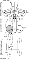
Figure 1.
Figure 1.
Schematic diagram of HD circuit and model of proposed information flow. The DTN is depicted with a diagonal line transecting it to represent the pars ventralis and pars dorsalis subdivisions of the DTN (Hayakawa and Zyo, 1990). The DTN receives prominent projections from both the nucleus prepositus hypoglossi, ipsilaterally, and the supragenual nucleus, contralaterally (shown on left only). Both are innervated by the medial vestibular nucleus and are therefore likely sources of AHV information to the DTN. These projections to the DTN compose one of what could be multiple idiothetic pathways of information about self-generated movement into the HD system. The ADN is reciprocally connected to the retrosplenial cortex, and to the postsubiculum. These projections may constitute an allothetic pathway through which cortically processed visual, motor, and mnemonic information could enter the HD system. The interconnections of the DTN and LMN may be sufficient to generate the characteristic activity of HD cells, whereas the idiothetic pathway updates that activity from moment to moment based on the animal's movement, and the allothetic pathway relates the representation of directional heading to salient spatial cues from the environment (Sharp et al., 2001a; Taube and Bassett, 2003). In the case of a unilateral lesion (left DTN; gray shading), only information reaching the right DTN from the contralateral supragenual nucleus and ipsilateral nucleus prepositus hypoglossi can reach either ADN via the LMN (lost connections shown as dotted lines). AHV input might still reach the left LMN via a direct projection from the supragenual nucleus (Biazoli et al., 2006). MVN, Medial vestibular nucleus; NPH, nucleus prepositus hypoglossi; PoS, postsubiculum; RSC, retrosplenial cortex; SGN, supragenual nucleus.
Figure 2.
Figure 2.
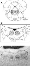
A, Location of the DTN. Schematic diagram of a coronal section at −9.16 mm posterior to bregma with DTN shaded bilaterally [modified from Paxinos and Watson (1998)]. B, Magnified view of the dorsal tegmental area. Enlargement of boxed area in A showing dorsal (TDD) and ventral (TDV) subdivisions of the DTN. Also visible are other structures most likely to be involved in DTN lesions: locus ceruleus (LC), laterodorsal tegmental nucleus (LDTg), mlf, and ventral tegmental nucleus (VTg). Also shown as landmarks are the mesencephalic trigeminal motoneurons (Me5). C, Photomicrograph of section falling approximately within the boxed area in A and B in a control rat showing the DTN (black outline) with its characteristic darkly stained ventral subdivision and the large motor trigeminal neurons visible laterally as landmarks.
Figure 3.
Figure 3.
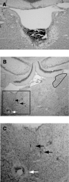
A, Complete DTN lesion. A coronal section at approximately −9.2 mm posterior to bregma from a lesioned rat shows complete loss of tissue in the immediate vicinity of the DTN and an area of damage that extends ventrally through the border of the tegmental gray area, with the motor trigeminal neurons still visible. B, Recording electrode path. Wire tracks through the ADN from the same rat shown in A reflect good sampling of cells within the ADN. Electrode wire tracks are visible within the ADN (black arrows), and a Prussian blue mark is visible below the ventral extent of the ADN (white arrow). Compression has distorted the ADN and neighboring tissue, but the dark cells of the ADN are still visible. Compare with the right ADN (black outline). C, Magnified view of electrode tracks showing boxed area in B.
Figure 4.
Figure 4.
Incomplete DTN lesions. A, Complete unilateral DTN lesion. The right DTN of this animal (rat 76) is visible fully intact (black outline) in this photomicrograph, yet this animal yielded no directionally modulated cells. The open space dorsal to the lesion site shows distortion of the tissue from downward pressure from the advancing electrode bundle. B, Incomplete DTN lesion. The right DTN of this animal (rat 75) is completely absent. Although tissue remains in the area of the left DTN (black outline), DTN neurons cannot be clearly identified. Recordings from this animal yielded two HD/AHV cells and six AHV-only cells.
Figure 5.
Figure 5.
Directional activity in rats with DTN lesions. Firing rate-by-head direction functions are shown for all cells that were classified as directionally modulated according to a Rayleigh test. Mean vector length (r) and level of significance are shown for each cell, and cells are ranked from left to right and top to bottom in order of their value for r. Note that the cells with the top four highest mean vector lengths are not noticeably different from normal HD cells and were classified as classic HD cells. The lower six cells were considered coarsely modulated by HD. Asterisks denote cells that were also modulated by AHV.
Figure 6.
Figure 6.
AHV in HD cells. A, Firing rate-by-head direction function for an ADN HD cell in a DTN-lesioned rat. B, Firing rate-by-AHV function for the same cell as A. Gray points indicate AHV samples taken from a 120° segment of the cell's directional firing range; black points indicate AHV samples taken from a 120° segment outside of the directional firing range. Gray and black points in B correspond to gray and black lines in A offset by vertical hash marks. Note that firing rate-by-AHV functions inside the directional firing range retain the symmetric shape of the function but are elevated at both baseline and peak velocities, indicating modulation by both AHV and HD. C, Firing rate-by-head direction function for an ADN HD cell in an unlesioned rat. D, Firing rate-by-AHV function for the same cell as C. Conventions are the same as for A and B. No AHV sensitivity is evident for this cell inside or outside of the directional firing range. Negative AHV values (left of 0) refer to clockwise directions; positive AHV values (right of 0) refer to counterclockwise directions.
Figure 7.
Figure 7.
AHV cells in a DTN-lesioned rat. Firing rate-by-AHV functions plotted for ADN cells recorded in DTN-lesioned rats. A, A negatively modulated cell similar to a small number of AHV cells in the DTN and LMN. B, A positively modulated cell that fires with head turns in either direction (type III). Note that firing rates are primarily correlated to AHV at velocities ≤100°/s in both clockwise and counterclockwise turn directions. C, An asymmetric cell that fires in the direction of contraversive head turn (type II). Negative AHV values (left of 0) refer to clockwise directions; positive AHV values (right of 0) refer to counterclockwise directions.
Figure 8.
Figure 8.
Place cells in a DTN-lesioned rat. A, Firing rate-by-place functions plotted for five hippocampal cells recorded in a DTN-lesioned rat (Table 1, rat 149). B, A place cell recorded when the white cue card was rotated +90°. As in intact animals, the place field followed the cue. C, Central plot shows firing rate by place for all head directions, as with a standard place cell analysis. The outer eight place–rate maps represent place-modulated activity when the rat's head is pointing in the direction indicated by the plot. For all plots, white pixels indicate that the animal did not visit the location, yellow pixels represent locations visited by the rat but at which the cell did not fire, and other pixels are assigned colors in ascending order according to their firing rate (orange, red, green, blue, and purple), with orange representing the lowest firing rates and purple the highest firing rates. See Materials and Methods for details.
Figure 9.
Figure 9.
A, Complete LMN lesion. A coronal section at approximately −4.5 mm posterior to bregma from a lesioned rat shows complete loss of tissue confined to the area of the mammillary nuclei, including both the LMN and medial mammillary nuclei, although a small patch of the medial mammillary nuclei remains visible in the center of the lesioned area. B, Recording electrode path. Wire tracks through the ADN from the same rat shown in A reflect good sampling of cells within the ADN. Electrode wire tracks are visible within the ADN and stria medullaris. Compare with the left ADN (black outline). C, Magnified view of electrode tracks showing boxed area in B; black arrows indicate electrode tracks, and the white arrow indicates Prussian blue mark.

Similar articles

Cited by

References

    1. Allen GV, Hopkins DA. Mamillary body in the rat: topography and synaptology of projections from the subicular complex, prefrontal cortex, and midbrain tegmentum. J Comp Neurol. 1989;286:311–336. - PubMed
    1. Allen GV, Hopkins DA. Topography and synaptology of mamillary body projections to the mesencephalon and pons in the rat. J Comp Neurol. 1990;301:214–231. - PubMed
    1. Bassett JP, Taube JS. Neural correlates for angular head velocity in the rat dorsal tegmental nucleus. J Neurosci. 2001;21:5740–5751. - PMC - PubMed
    1. Bassett JP, Taube JS. Head direction signal generation: ascending and descending information streams. In: Wiener SI, Taube JS, editors. Head direction cells and the neural mechanisms of spatial orientation. Cambridge, MA: MIT; 2005. pp. 83–109.
    1. Batschelet E. New York: Academic; 1981. Circular statistics in biology.

Publication types