

Trisomy 19 ependymoma, a newly recognized genetico-histological association, including clear cell ependymoma
- PMID: 17626628
- PMCID: PMC1950527
- DOI: 10.1186/1476-4598-6-47
Trisomy 19 ependymoma, a newly recognized genetico-histological association, including clear cell ependymoma
Abstract
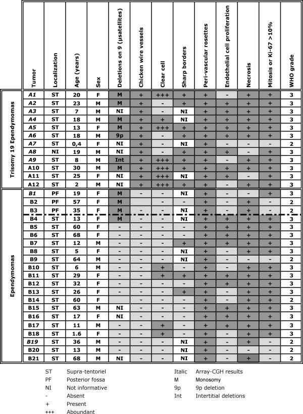
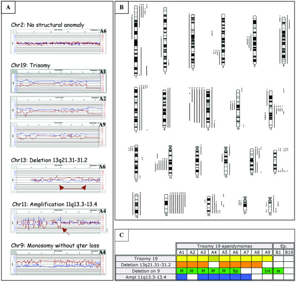
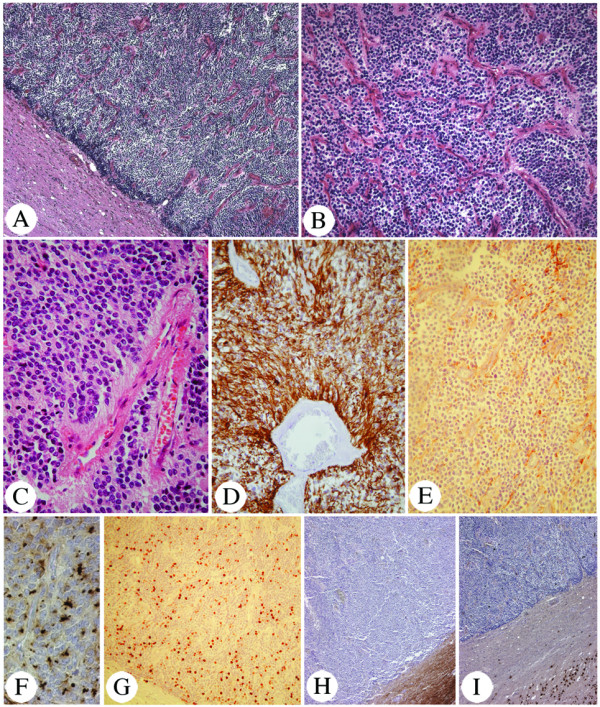
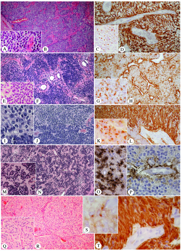
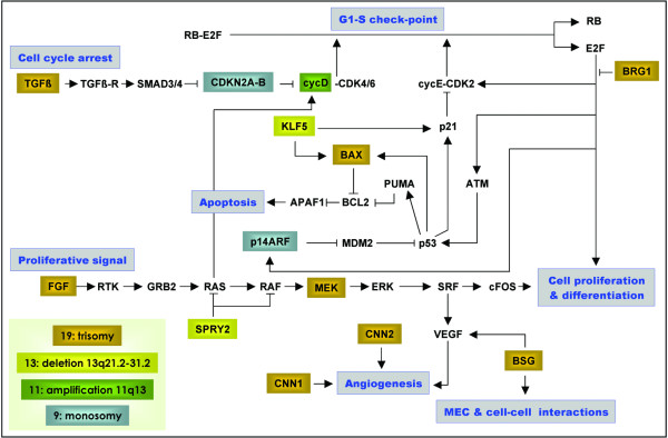
Ependymal tumors constitute a clinicopathologically heterogeneous group of brain tumors. They vary in regard to their age at first symptom, localization, morphology and prognosis. Genetic data also suggests heterogeneity. We define a newly recognized subset of ependymal tumors, the trisomy 19 ependymoma. Histologically, they are compact lesions characterized by a rich branched capillary network amongst which tumoral cells are regularly distributed. When containing clear cells they are called clear cell ependymoma. Most trisomy 19 ependymomas are supratentorial WHO grade III tumors of the young. Genetically, they are associated with trisomy 19, and frequently with a deletion of 13q21.31-31.2, three copies of 11q13.3-13.4, and/or deletions on chromosome 9. These altered chromosomal regions are indicative of genes and pathways involved in trisomy 19 ependymoma tumorigenesis. Recognition of this genetico-histological entity allows better understanding and dissection of ependymal tumors.
Figures
Similar articles
-
Pediatric supratentorial ependymomas show more frequent deletions on chromosome 9 than infratentorial ependymomas: a microsatellite analysis.Cancer Genet Cytogenet. 2009 Jun;191(2):90-6. doi: 10.1016/j.cancergencyto.2009.02.010. Cancer Genet Cytogenet. 2009. PMID: 19446744
-
Human ependymomas reveal frequent deletions on chromosomes 6 and 9.Acta Neuropathol. 2003 Oct;106(4):357-62. doi: 10.1007/s00401-003-0739-5. Epub 2003 Jul 24. Acta Neuropathol. 2003. PMID: 12898154
-
Candidate genes on chromosome 9q33-34 involved in the progression of childhood ependymomas.J Clin Oncol. 2009 Apr 10;27(11):1884-92. doi: 10.1200/JCO.2007.15.4195. Epub 2009 Mar 16. J Clin Oncol. 2009. PMID: 19289631
-
[Diagnostic and prognostic values of 1p and 19q deletions in adult gliomas: critical review of the literature and implications in daily clinical practice].Rev Neurol (Paris). 2008 Jun-Jul;164(6-7):595-604. doi: 10.1016/j.neurol.2008.04.002. Epub 2008 May 21. Rev Neurol (Paris). 2008. PMID: 18565359 Review. French.
-
Pediatric ependymoma: biological perspectives.Mol Cancer Res. 2009 Jun;7(6):765-86. doi: 10.1158/1541-7786.MCR-08-0584. Epub 2009 Jun 16. Mol Cancer Res. 2009. PMID: 19531565 Review.
Cited by
-
Chromosome 7 and 19 trisomy in cultured human neural progenitor cells.PLoS One. 2009 Oct 29;4(10):e7630. doi: 10.1371/journal.pone.0007630. PLoS One. 2009. PMID: 19898616 Free PMC article.
-
Supratentorial clear cell ependymomas with branching capillaries demonstrate characteristic clinicopathological features and pathological activation of nuclear factor-kappaB signaling.Neuro Oncol. 2016 Jul;18(7):919-27. doi: 10.1093/neuonc/now025. Epub 2016 Mar 15. Neuro Oncol. 2016. PMID: 26984744 Free PMC article.
-
Supra- and infratentorial pediatric ependymomas differ significantly in NeuN, p75 and GFAP expression.J Neurooncol. 2013 Apr;112(2):191-7. doi: 10.1007/s11060-013-1062-1. Epub 2013 Jan 31. J Neurooncol. 2013. PMID: 23371454
-
Intracranial ependymoma associated with multiple endocrine neoplasia type 1.J Endocrinol Invest. 2010 May;33(5):353-6. doi: 10.1007/BF03346599. Epub 2010 Feb 5. J Endocrinol Invest. 2010. PMID: 20142633 No abstract available.
-
Supratentorial "vascular" variant of ependymoma: a lesser known morphologic variant and a diagnostic pitfall.Childs Nerv Syst. 2016 Sep;32(9):1569-71. doi: 10.1007/s00381-016-3158-3. Epub 2016 Jul 8. Childs Nerv Syst. 2016. PMID: 27392446 No abstract available.
References
-
- Kleihues P, Cavenee WK, (Eds) Pathology & Genetics. Tumours of the Nervous System. Lyon: IARC Press; 2000.
Publication types
MeSH terms
LinkOut - more resources
Full Text Sources
Medical
