Noninvasive monitoring of myocardial function after surgical and cytostatic therapy in a peritoneal metastasis rat model: assessment with tissue Doppler and non-Doppler 2D strain echocardiography
- PMID: 17626632
- PMCID: PMC1965460
- DOI: 10.1186/1476-7120-5-23
Noninvasive monitoring of myocardial function after surgical and cytostatic therapy in a peritoneal metastasis rat model: assessment with tissue Doppler and non-Doppler 2D strain echocardiography
Abstract
Objective: We sought to evaluate the impact of different antineoplastic treatment methods on systolic and diastolic myocardial function, and the feasibility estimation of regional deformation parameters with non-Doppler 2D echocardiography in rats.
Background: The optimal method for quantitative assessment of global and regional ventricular function in rats and the impact of complex oncological multimodal therapy on left- and right-ventricular function in rats remains unclear.
Methods: 90 rats after subperitoneal implantation of syngenetic colonic carcinoma cells underwent different onclogical treatment methods and were diveded into one control group and five treatment groups (with 15 rats in each group): group 1 = control group (without operation and without medication), group 2 = operation group without additional therapy, group 3 = combination of operation and photodynamic therapy, group 4 = operation in combination with hyperthermic intraoperative peritoneal chemotherapy with mitomycine, and group 5 = operation in combination with hyperthermic intraoperative peritoneal chemotherapy with gemcitabine, group 6 = operation in combination with taurolidin i.p. instillation. Echocardiographic examination with estimation of wall thickness, diameters, left ventricular fractional shortening, ejection fraction, early and late diastolic transmitral and myocardial velocities, radial and circumferential strain were performed 3-4 days after therapy.
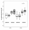
Results: There was an increase of LVEDD and LVESD in all groups after the follow-up period (P = 0.0037). Other LV dimensions, FS and EF as well as diastolic mitral filling parameters measured by echocardiography were not significantly affected by the different treatments. Values for right ventricular dimensions and function remained unchanged, whereas circumferential 2D strain of the inferior wall was slightly, but significantly reduced under the treatment (-18.1 +/- 2.5 before and -16.2 +/- 2.9 % after treatment; P = 0.001) without differences between the single treatment groups.
Conclusion: It is feasible to assess dimensions, global function, and regional contractility with echocardiography in rats under different oncological therapy. The deformation was decreased under overall treatment without influence by one specific therapy. Therefore, deformation assessment with non-Doppler 2D strain echocardiography is more sensitive than conventional echocardiography for assessing myocardial dysfunction in rats under oncological treatment.
Figures





References
-
- Vogel M, Schmidt MR, Kristiansen SB, Cheung M, White PA, Sorensen K, Redington AN. Validation of myocardial acceleration during isovolumic contraction as a novel noninvasive index of right ventricular contractility. Comparison with ventricular pressure-volume relations in an animal model. Circulation. 2002;105:1693–1699. doi: 10.1161/01.CIR.0000013773.67850.BA. - DOI - PubMed
-
- Borges AC, Knebel F, Eddicks S, Panda A, Schattke S, Witt C, Baumann G. Right ventricular function assessed by two-dimensional strain and tissue Doppler echocardiography in patients with pulmonary arterial hypertension and effect of vasodilator therapy. Am J Cardiol. 2006;98:530–534. doi: 10.1016/j.amjcard.2006.02.060. - DOI - PubMed
-
- Leitman M, Lysyansky P, Sidenko S, Shir W, Peleg E, Binenbaum M, Kaluski E, Karkover R, Vered Z. Two-dimensional strain a novel software for real-time quantitative echocardiographic assessment of myocardial function. J Am Soc Echocardiogr. 2004;17:1021–1029. doi: 10.1016/j.echo.2004.06.019. - DOI - PubMed
MeSH terms
Substances
LinkOut - more resources
Full Text Sources
Medical

