. 2007 Sep 1;74(5):768-79.
doi: 10.1016/j.bcp.2007.06.003. Epub 2007 Jun 7.

Role of adenosine A3 receptors on CA1 hippocampal neurotransmission during oxygen-glucose deprivation episodes of different duration

Affiliations

Role of adenosine A3 receptors on CA1 hippocampal neurotransmission during oxygen-glucose deprivation episodes of different duration

Anna Maria Pugliese et al. Biochem Pharmacol. .

Abstract

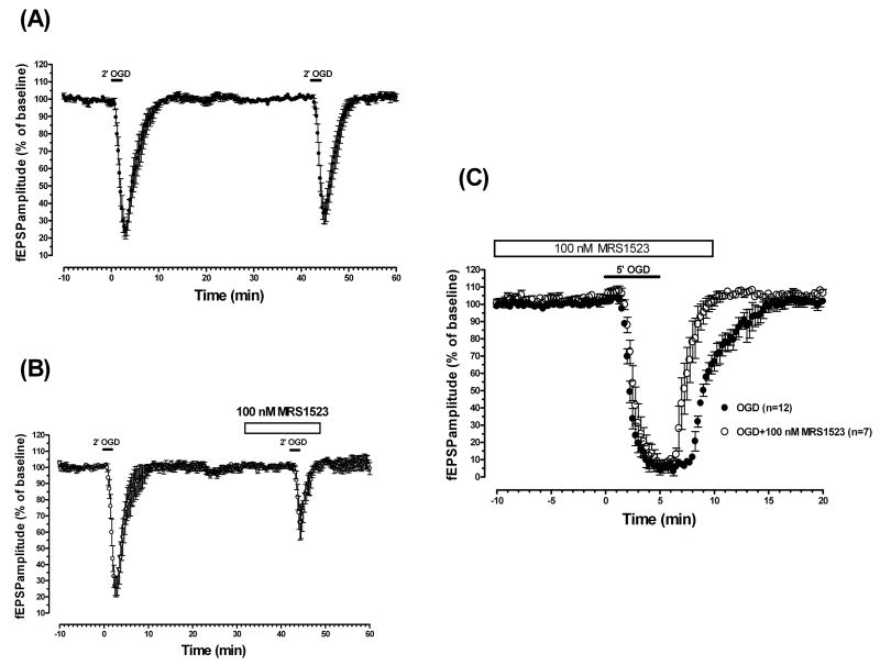
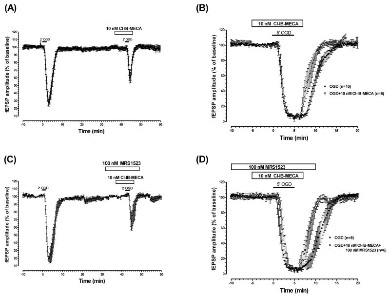
The role of adenosine A3 receptors in synaptic transmission under severe (7 min) and shorter (2-5 min) ischemic conditions, obtained by oxygen and glucose deprivation (OGD), was investigated in rat hippocampal slices. The effects of selective A3 agonists or antagonists were examined on field excitatory postsynaptic potentials (fEPSPs) extracellularly recorded at the dendritic level of the CA1 pyramidal region. The novel, selective A3 antagonist LJ1251 ((2R,3R,4S)-2-(2-chloro-6-(3-iodobenzylamino)-9H-purin-9-yl)tetrahydrothiophene-3,4-diol, 0.1-10 nM) protected hippocampal slices from irreversible fEPSP depression induced by severe OGD and prevented or delayed the appearance of anoxic depolarization. Similar results were obtained when severe OGD was carried out with a long, receptor-desensitizing exposure to various selective A3 agonists: 5'-N-methylcarboxamidoadenosine derivatives Cl-IB-MECA (N6-(3-iodobenzyl)-2-chloro), VT72 (N6-methoxy-2-phenylethynyl), VT158 (N6-methoxy-2-phenylethynyl), VT160 (N6-methoxy-2-(2-pyridinyl)-ethynyl), and VT163 (N6-methoxy-2-p-acetylphenylethynyl) and AR132 (N6-methyl-2-phenylethynyladenosine). The selective A3 antagonist MRS1523 (3-propyl-6-ethyl-5-[(ethylthio)carbonyl]-2-phenyl-4-propyl-3-pyridine carboxylate, 100 nM) reduced fEPSP depression evoked by 2-min OGD and induced a faster recovery of fEPSP amplitude after 5-min OGD. Similar results were obtained for 2- or 5-min OGD applied in the presence of each of the A3 agonists tested. Shorter exposure to A3 agonists significantly delayed the recovery of fEPSP amplitude after 5-min OGD. This indicates that A3 receptors, stimulated by selective A3 agonists, undergo desensitization during OGD. It is inferred that CA1 hippocampal A3 receptors stimulated by adenosine released during brief ischemia (2 and 5 min) might exert A1-like protective effects on neurotransmission. Severe ischemia would transform the A3 receptor-mediated effects from protective to injurious.


Figures

Fig. 1
Fig. 1
The selective A3 adenosine receptor antagonists, LJ1251 and MRS1523, minimized AD and protected CA1 hippocampus from irreversible fEPSP depression induced by 7-min OGD. (A) AD, was recorded as the negative d.c. shift in response to 7-min OGD (solid bar) in the absence (n = 13, left panel), and in the presence of 10 nM LJ1251 (open bar, n = 5, middle panel) or 100 nM MRS1523 (open bar, n = 5, right panel). Note that both the A3 antagonists LJ1251 and MRS1583 completely prevented AD appearance. (B) Graphs show the time course of 7-min OGD (solid bar) effect on fEPSP amplitude, expressed as percentage of baseline, in untreated OGD slices (filled circles, mean ± S.E.M., n = 13), in the presence of 10 nM LJ1251 (open circles, mean ± S.E.M., n = 5) and 100 nM MRS1523 (filled triangles, mean ± S.E.M., n=5). Solid bars indicate the duration of OGD. (C) Concentration-response curves of LJ1251. The graph shows the fitting curve of concentration-dependent effects of LJ1251 on fEPSP recovery after 7-min OGD recorded in hippocampal slices at 15-min reperfusion in normal, oxygenated aCSF. Data (mean ± S.E.M., n = 3 in 0.001 nM LJ1251, n = 3 in 0.01 nM, n = 3 in 0.1 nM, n = 4 in 5 nM, n = 5 in 10 nM) are expressed as percentage of baseline values. *P < 0.01, one-way ANOVA, Newman-Keuls multiple-comparison post hoc test, versus 0.001 and 0.01 nM LJ1251-treated slices.
Fig. 2
Fig. 2
The selective A3 adenosine agonist Cl-IB-MECA protects hippocampal slices from the irreversible fEPSP depression induced by 7-min OGD. (A) AD was recorded as the negative d.c. shift in response to 7-min OGD (solid bars) in the absence (n = 8) and in the presence of 10 nM Cl-IB-MECA (open bar, n = 8). The agonist prevented AD appearance in 5 slices and delayed it in the remaining 3 slices. (B) Time course of fEPSP amplitude before, during 7-min OGD, and after reperfusion in normal oxygenated aCSF in the absence and in the presence of 10 nM Cl-IB-MECA (open bar). Data (mean ± S.E.M., n = 16 in untreated OGD slices, n = 15 in Cl-IB-MECA) are expressed as percentage of baseline values. Cl-IB-MECA was applied 5 min before, during, and 2 min after OGD. (C) Columns in the graph summarize the effects of different A3 agonists on fEPSP recovery after 7- min OGD. *P < 0.05, one-way ANOVA followed by Newman-Keuls multiple-comparison post hoc test versus untreated OGD slices (D) Each graph shows the time course of fEPSP amplitude before anf after 7-min OGD. Data (mean ± S.E.M., n = 3 for each experimental group) are expressed as percentage of baseline values. Note that in a group of slices OGD was applied 20 min after the end of Cl-IB-MECA superfusion (open bar, 10 nM, 15 min). A complete recovery of fEPSP amplitude was obtained only in the slices in which OGD was applied after Cl-IB-MECA application. Solid bars indicate the duration of OGD.
Fig. 3
Fig. 3
The selective A3 adenosine receptor antagonist MRS1523 reduces fEPSP depression induced by 2-min OGD and induced a faster recovery of fEPSP amplitude after 5-min OGD. (A) Time course of fEPSP amplitude before, during, and after the application of two consecutive ischemic insults of 2-min duration. The second 2-min OGD, administered 40 min after the end of the first period, elicited a comparable depression of synaptic potentials (P > 0.05, evaluated by t-paired two-tailed Student's t test). Data (mean ± S.E.M., n = 13) are expressed as a percentage of baseline values recorded before the first OGD period. (B) Time course of fEPSP amplitude changes elicited by 2-min OGD in the absence and in the presence of MRS1523. Data (mean ± S.E.M., n = 10) are expressed as a percentage of baseline values. MRS1523 (100 nM) was applied 10 min before, during, and 5 min after OGD, as indicated by the open bar. Note that in the presence of MRS1523, the fEPSP depression induced by OGD was significantly lower than that obtained in the absence of the drug (P < 0.05, paired two-tailed Student's t test). (C) Time course of fEPSP amplitude changes elicited by 5-min OGD in the absence (n = 12) and in the presence of MRS1523 (n = 7). Data (mean ± S.E.M.) are expressed as a percentage of baseline values recorded before the respective OGD periods. MRS1523 (100 nM), applied 10 minutes before, during, and 5 minutes after OGD application induced a faster fEPSP recovery after OGD application. Solid bars indicate the duration of OGD.
Fig. 4
Fig. 4
The selective A3 adenosine receptor agonist Cl-IB-MECA decreased fEPSP depression induced by 2-min OGD and induced a faster recovery of fEPSP amplitude after 5-min OGD. (A) Time-course of fEPSP amplitude changes elicited by 2-min OGD in the absence and in the presence of Cl-IB-MECA (10 nM; n = 13). Data (mean ± S.E.M.) are expressed as a percentage of respective baseline values recorded before the respective OGD periods. The solid bar indicates the duration of OGD. As indicated by the open bar, the A3 agonist was applied 5 min before, during, and 2 min after OGD. Note that in the presence of Cl-IB-MECA, the fEPSP depression induced by 2-min OGD was significantly lower than that obtained in the absence of the drug (P < 0.05, paired two-tailed Student's t test). (B) Time course of fEPSP amplitude changes elicited by 5 min OGD in the absence (n = 10) and presence of Cl-IB-MECA (10 nM, n = 6). Data (mean ± S.E.M.) are expressed as a percentage of baseline values recorded before the respective OGD periods. The A3 agonist, applied 5 min before, during, and 2 min after OGD (open bar) induced a faster fEPSP recovery after 5 min OGD. (C) Time course of fEPSP amplitude changes elicited by 2-min OGD in the absence and presence of Cl-IB-MECA (10 nM) in combination with MRS1523 (100 nM). The antagonist was always superfused 5 min before the agonist; coapplied with the agonist 5 min before, during, and 2 min after OGD; and then superfused alone for 3 min. Data (mean ± S.E.M., n = 7) are expressed as a percentage of the respective baseline values. Note that in the presence of Cl-IB-MECA and MRS1523, the fEPSP depression induced by 2-min OGD was significantly lower than (P < 0.05, paired two-tailed Student's t test) that obtained in the absence of the drugs. (D) Time course of fEPSP amplitude changes elicited by 5 min OGD in the absence (n = 8) and presence of Cl-IB-MECA (10 nM) in combination with MRS1523 (100 nM) (n = 6). As indicated by open bars, the antagonist was always superfused 5 min before the agonist; coapplied with the agonist 5 min before, during and 2-min after OGD; and then superfused alone for 3 min. Data (mean ± S.E.M.) are expressed as a percentage of respective baseline values. Solid bars indicate the duration of OGD.
Fig. 5
Fig. 5
Short application Cl-IB-MECA did not modify fEPSP depression induced by 2-min OGD, but it significantly delays fEPSP recovery after 5-min OGD. (A) Time course of fEPSP amplitude changes elicited by 2-min OGD in the absence and in the presence of Cl-IB-MECA (10 nM, n = 7). Data (mean ± S.E.M.) are expressed as a percentage of respective baseline values recorded before the respective OGD periods. As indicated by the open bar, the A3 agonist was applied for only 2 min, during OGD. Note that short application of Cl-IB-MECA did not induce significant change (P > 0.05, paired two-tailed Student's t test) in the fEPSP depression induced by 2-min OGD. (B) Time course of fEPSP amplitude changes elicited by 5-min OGD in the absence (n = 20) and in the presence of Cl-IB-MECA (10 nM, n = 10). Data (mean ± S.E.M.) are expressed as a percentage of respective baseline values recorded before the respective OGD periods. As indicated by the open bar, the A3 agonist was applied only for 2 min, at the end of 5-min OGD episodes. Note that short application of Cl-IB-MECA delayed fEPSP recovery after 5 min OGD. (C) Columns in the graph summarize the average time of fEPSP amplitude recovery (mean ± S.E.M.) after 5-min OGD in untreated OGD slices and during long or short application of Cl-IB-MECA. *P < 0.05, one-way ANOVA followed by Newman-Keuls multiple-comparison post hoc test versus untreated OGD slices; § P < 0.05, one-way ANOVA followed by Newman-Keuls multiple-comparison post hoc test versus all experimental groups. Solid bars indicate the duration of OGD.

Similar articles

Cited by

References

    1. Fredholm BB, IJzerman AP, Jacobson KA, Klotz KN, Linden J. International Union of Pharmacology. XXV. Nomenclature and classification of adenosine receptors. Pharmacol Rev. 2001;53:527–552. - PMC - PubMed
    1. Zhou QY, Li C, Olah ME, Johnson RA, Stiles GL, Civelli O. Molecular cloning and characterization of an adenosine receptor: the A3 adenosine receptor. Proc Natl Acad Sci USA. 1992;89:7432–7436. - PMC - PubMed
    1. Dixon AK, Gubitz AK, Sirinathsinghji DJ, Richardson PJ, Freeman TC. Tissue distribution of adenosine receptor mRNAs in the rat. Br J Pharmacol. 1996;118:1461–1468. - PMC - PubMed
    1. Diaz-Hernandez M, Pereira MF, Pintor J, Cunha RA, Ribeiro JA, Miras-Portugal MT. Modulation of the rat hippocampal dinucleotide receptor by adenosine receptor activation. J Pharmacol Exp Ther. 2002;301:441–450. - PubMed
    1. Lopes LV, Rebola N, Pinheiro PC, Richardson PJ, Oliveira CR, Cunha RA. Adenosine A3 receptors are located in neurons of the rat hippocampus. Neuroreport. 2003;14:1645–1648. - PubMed

Publication types