. 2007 Jul 15;21(14):1731-46.
doi: 10.1101/gad.1556607. Epub 2007 Jul 12.

Discovery of an oncogenic activity in p27Kip1 that causes stem cell expansion and a multiple tumor phenotype

Affiliations

Discovery of an oncogenic activity in p27Kip1 that causes stem cell expansion and a multiple tumor phenotype

Arnaud Besson et al. Genes Dev. .

Abstract

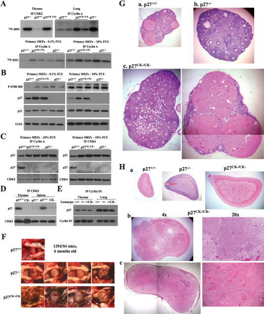
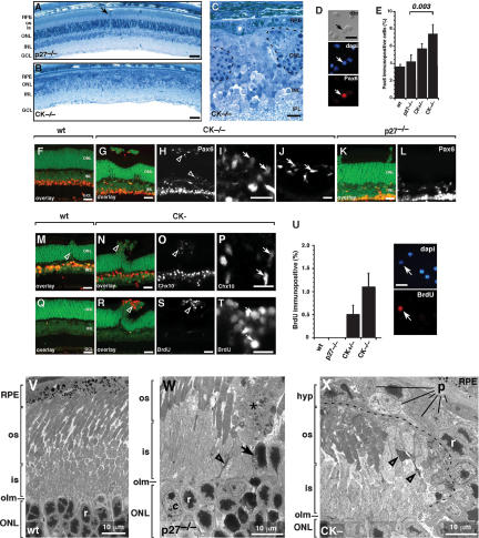
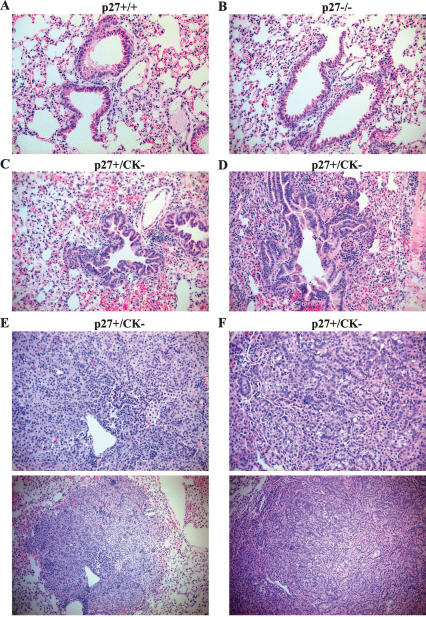
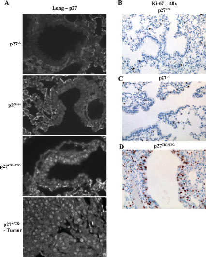
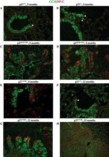
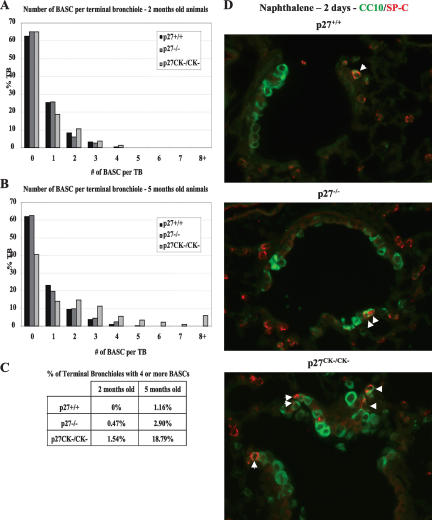
The cell cycle inhibitor p27Kip1 also has cyclin-cyclin-dependent kinase (CDK)-independent functions. To investigate the significance of these functions in vivo, we generated a knock-in mouse in which four amino acid substitutions in the cdkn1b gene product prevent its interaction with cyclins and CDKs (p27CK-). In striking contrast to complete deletion of the cdkn1b gene, which causes spontaneous tumorigenesis only in the pituitary, the p27CK- protein dominantly caused hyperplastic lesions and tumors in multiple organs, including the lung, retina, pituitary, ovary, adrenals, spleen, and lymphomas. Moreover, the high incidence of spontaneous tumors in the lung and retina was associated with amplification of stem/progenitor cell populations. Therefore, independently of its role as a CDK inhibitor, p27Kip1 promoted stem cell expansion and functioned as a dominant oncogene in vivo. Thus, the p27CK- mouse unveils a dual role for p27 during tumorigenesis: It is a tumor suppressor by virtue of its cyclin-CDK regulatory function, and also an oncogene through a cyclin-CDK-independent function. This may explain why the cdkn1b gene is rarely inactivated in human tumors, and the p27CK- mouse in which the tumor suppressor function is lost but the cyclin-CDK-independent-oncogenic-function is maintained may represent a more faithful model for the widespread role of p27 misregulation in human cancers than the p27 null.


Figures

Figure 1.
Figure 1.
The p27CK allele does not regulate cyclin–CDK complexes and causes multiple organs hyperplasia. (A) Comparison of cyclin A and CDK2-associated kinase activities in lung, thymus, and primary MEFs (either serum-starved for 48 h [0.1% FCS] or exponentially growing [10% FCS]) derived from wild-type, p27−/−, and p27CK/CK mice. (B) Increased phospho-Rb levels in p27CK/CK and p27−/− primary fibroblasts. Serum-starved or exponentially growing MEFs were probed sequentially for Phospho-Ser780-Rb (a CDK phosphorylation site), p27, p21, and Grb2 for loading control. (C) p27CK does not interact with cyclins or CDKs and does not prevent p21 from binding to them. Exponentially growing primary MEF extracts were immunoprecipitated for cyclin A and CDK4 and were probed for p21, p27, and CDK2 (in cyclin A immunoprecipitates) and CDK4 (in CDK4 immunoprecipitates). (D) p27CK does not bind to CDK2 in thymus and spleen. CDK2 immunoprecipitates from thymus and spleen extracts from wild-type, p27-null, and p27CK mice were probed for p27 and CDK2. (E) p27CK in p27+/CK mice does not prevent the remaining wild-type p27 from interacting with cyclin D1. Cyclin D1 immunoprecipitates from thymus and lung of wild-type, p27+/−, and p27+/CK mice were probed for p27 and cyclin D1. (F) Accelerated pituitary tumorigenesis in p27CK/CK mice compared with wild-type and p27−/− animals at 6 mo of age. (G) H&E staining of ovaries from 5- to 6-mo-old mice (magnification, 4×). (Panel a) Wild-type ovary showing normal follicle maturation. (Panel b) Ovaries from p27−/− mice are enlarged and lack corpora lutea. (Panel c) Twenty-five percent of p27CK ovaries form corpora lutea, which are often grossly enlarged (as shown in right panel). (H) Spontaneous pheochromocytoma in p27CK/CK mice. (Panel a) H&E-stained sections of adrenal glands from 6-mo-old wild-type, p27−/−, and p27CK/CK mice (magnification, 4×). (Panels b,c) Pheochromocytomas developing in 12-mo-old (panel b) and 14-mo-old (panel c) p27CK/CK mice.
Figure 2.
Figure 2.
Hyperplasia in p27CK retinae. (AC) Toluidine blue-stained 1-μm plastic sections of adult p27−/− (A) and p27CK (B) retinae. The ONL is severely disrupted and hypocellular, and the inner and outer segments are absent in the p27CK. (C) Higher-magnification view of the p27CK retina, showing hyperplasia of cells with an immature morphology invading the ONL (dashed line). (D,E) Dissociated cell scoring of adult retinae for Pax6 immunoreactivity. (D) Example of a Pax6 immunopositive cell from the p27CK retina. (E) Five-hundred cells were scored in duplicate from three independent animals, and the mean and standard deviation are presented. There was a statistically significant increase in Pax6 immunopositive cells in the p27CK retina as compared with the p27-null retina (P = 0.003). (FL) Immunostained vibratome sections from wild-type, p27-null, and p27CK retinae showing the pattern of Pax6 immunoreactive cells. Lower levels of Pax6 are consistent with retinal progenitor cells as compared with the high levels seen in differentiated amacrine cells in the INL. Ectopic Pax6 immunopositive cells were present at the apical edge of the retina (magnification in I) as well as the basal edge of the ONL (magnification in J). (K,L) p27-null retinae do not show this pattern of Pax6 expression associated with the mild form of retinal dysplasia seen in these retinae. (M) Chx10 is expressed in retinal progenitor cells and differentiated bipolar cells. (N,O) In the p27CK retinae, Chx10 is expressed in the bipolar cells as well as ectopic cells extending apically to the outer limiting membrane (olm). (P) A magnified view of the cells indicated by the open arrowhead in O. The ectopic Chx10-expressing cells are not found in the p27-null retinae (shown in M). (QU) Adult retinae were labeled for 1 h with 10 μM BrdU, and then retinal sections were immunostained for BrdU along with dissociated retinal cells from the same animal (shown in U). (Q) Wild-type adult retinae show now BrdU immunopositive nuclei. (R,S) However, p27CK retinae had BrdU immunopositive nuclei associated with the retinal dysplasia located through the ONL (open arrowhead). (T) A higher-magnification view of the BrdU immunopositive cells shown in R and S. (VX) Transmission electron microscopy of wild-type (V), p27-null (W), and p27CK (X) retinae. (V) In the wild-type retina, the outer limiting membrane (olm) separates the photoreceptor cell bodies from the photoreceptor inner segments (is) and outer segments (os). (W) In the p27-deficient retinae, there is an occasional cell that has been displaced apically beyond the outer limiting membrane (olm; arrow). These cells have the morphological features of rod photoreceptors. Such displaced photoreceptors are also associated with cellular debris in the dysplastic lesion (*). There is also evidence for photoreceptor cell death in the inner segments of photoreceptors in and around the retinal dysplasia in p27-null retinae (open arrowhead). (X) In the p27CK retinae, hyperplastic lesions of cells with morphological features of progenitor cells (p) migrate apically and separate the photoreceptors (dashed line) from their overlying RPE cells. It is likely that this leads to photoreceptor cell death (open arrowheads). (r) Rod photoreceptor, (p) progenitor cell, (is) inner segments, (os) outer segments, (RPE) retinal pigment epithelium, (ONL) outer nuclear layer, (INL) inner nuclear layer, (GCL) ganglion cell layer, (olm) outer limiting membrane. Bars: A,B, 50 μm; all others, 10 μm.
Figure 3.
Figure 3.
Spontaneous lung tumor formation in p27CK mice. Representative images of H&E-stained lung sections of wild-type (A), p27−/− (B), and p27+/CK (CF) mice (magnification, 20×); in the bottom panels in E and F, magnification is 4×. Panels CF show the progressive changes occurring in the bronchioalveolar epithelium of p27CK mice as follows: hyperplasia (C), early dysplastic changes (D), lung adenoma (E), and adenocarcinoma (F).
Figure 4.
Figure 4.
DIP and adenocarcinomas masquerading as DIP in p27CK mice. (A) Pictures of lungs dissected from p27CK/CK and p27+/CK mice showing the typical discrete adenomas or adenocarcinomas (left panel) and the diffuse lesions caused by the DIP or DIP-like adenocarcinomas (right panel). (B, top panels) H&E-stained sections showing normal lung structure in a wild-type and DIP in a p27+/CK animal (magnification, 20×). (Bottom panels) Immunohistochemistry with the pan-macrophage marker F4/80 showing the absence of staining in the wild-type lung and abundant macrophage infiltration in the p27CK/CK lung (magnification, 40×). (C) The top panel shows a DIP-like lesion in a p27+/CK lung section stained with H&E. The bottom panels show a consecutive section of the same lung stained with a pan-keratin antibody to stain the bronchioalveolar epithelium, as shown in a healthy portion of the section (middle picture). (Bottom picture) The DIP-like region of the section revealed a keratin-positive carcinoma (magnification, 20×).
Figure 5.
Figure 5.
Cytoplasmic localization of p27 and dysregulated proliferation in lung epithelium in mice carrying the p27CK allele. (A) Paraffin-embedded lung sections from p27−/−, p27+/+, p27CK/CK, and p27+/CK mice were stained for p27. All images were acquired using the same exposure time and settings, with a 60× lens. (B–D). Sections of paraffin-embedded lungs from 5-mo-old wild-type (B), p27−/− (C), and p27CK/CK (D) mice were stained with a rabbit monoclonal antibody to Ki-67.
Figure 6.
Figure 6.
p27CK causes amplification of the BASC pool. Paraffin-embedded lung sections were stained for CCSP/CC10 (goat anti-CC10, green) and SP-C (rabbit anti-SP-C, red) and merged images are shown. All images were acquired using a 60× objective, except for the image in H, which was acquired with a 20× lens. In A, B, and F, BASCs are indicated with an arrowhead. The individual images as well as the fields with Hoescht-stained nuclei for each panel are provided in Supplementary Figure 7A–H.
Figure 7.
Figure 7.
Increased BASC number in p27CK/CK mice. (AC) The number of BASC per TB was determined in 2-mo-old (A) and 5-mo-old (B) wild-type, p27−/−, and p27CK/CK mice. (C) Summary of the percentage of TB having four or more BASCs for each genotype. (D) BASCs are resistant to naphthalene injury. Immunofluorescence for CCSP/CC10 (green) and SP-C (red) of lung sections collected 48 h post-naphthalene injection. Images were acquired with a 60× lens. The individual images as well as the fields with Hoescht-stained nuclei for each panel are provided in Supplementary Figure 8A–C.

Comment in

Similar articles

Cited by

References

    1. Ayvazian L.F. Desquamative interstitial pneumonia. A systemic disease and precancerous lesion? J. Med. Soc. N. J. 1974;71:115–119. - PubMed
    1. Besson A., Yong V.W., Yong V.W. Involvement of p21(Waf1/Cip1) in protein kinase C α-induced cell cycle progression. Mol. Cell. Biol. 2000;20:4580–4590. - PMC - PubMed
    1. Besson A., Assoian R.K., Roberts J.M., Assoian R.K., Roberts J.M., Roberts J.M. Regulation of the cytoskeleton: An oncogenic function for CDK inhibitors? Nat. Rev. Cancer. 2004a;4:948–955. - PubMed
    1. Besson A., Gurian-West M., Schmidt A., Hall A., Roberts J.M., Gurian-West M., Schmidt A., Hall A., Roberts J.M., Schmidt A., Hall A., Roberts J.M., Hall A., Roberts J.M., Roberts J.M. p27Kip1 modulates cell migration through the regulation of RhoA activation. Genes & Dev. 2004b;18:862–876. - PMC - PubMed
    1. Besson A., Gurian-West M., Chen X., Kelly-Spratt K.S., Kemp C.J., Roberts J.M., Gurian-West M., Chen X., Kelly-Spratt K.S., Kemp C.J., Roberts J.M., Chen X., Kelly-Spratt K.S., Kemp C.J., Roberts J.M., Kelly-Spratt K.S., Kemp C.J., Roberts J.M., Kemp C.J., Roberts J.M., Roberts J.M. A pathway in quiescent cells that controls p27Kip1 stability, subcellular localization and tumor suppression. Genes & Dev. 2006;20:47–64. - PMC - PubMed

Publication types

MeSH terms

Substances

LinkOut - more resources