

Activity in the human amygdala corresponds to early, rather than late period autonomic responses to a signal for shock
- PMID: 17626906
- PMCID: PMC1934343
- DOI: 10.1101/lm.632007
Activity in the human amygdala corresponds to early, rather than late period autonomic responses to a signal for shock
Abstract
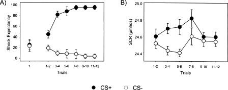
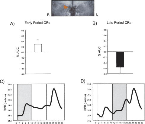
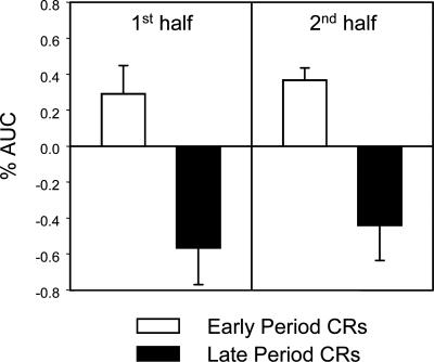
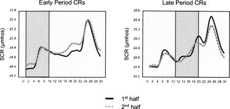
Laboratory animal and human subject studies report that the amygdala is a critical brain structure that supports the acquisition and expression of conditional fear. Recent functional neuroimaging studies in humans have reported that activity in this region is closely related to the behavioral expression of conditional skin conductance responses (SCR). However, SCR waveforms following conditional stimulus (CS) presentation contain both early period and late period responses that may differ with respect to underlying central processes. It is not known whether amygdala activity corresponds to the expression of early conditional responses (CRs) that occur shortly following CS onset or late CRs that closely precede UCS onset. The present study used event-related functional magnetic resonance imaging and concurrent skin conductance measurements to determine whether amygdala activity is more closely related to the expression of early or late period CRs. Increased amygdala activity was detected during the formation of early, but not late period CRs. Additionally, this pattern of amygdala activity did not dissipate, but persisted into late stages of the experiment. These findings are consistent with the idea that amygdala responding is critically involved in the generation of CRs formed shortly following CS onset.
Figures
Similar articles
-
Human amygdala activity during the expression of fear responses.Behav Neurosci. 2006 Dec;120(6):1187-95. doi: 10.1037/0735-7044.120.5.1187. Behav Neurosci. 2006. PMID: 17201461
-
Altered cingulate and amygdala response towards threat and safe cues in attention deficit hyperactivity disorder.Psychol Med. 2014 Jan;44(1):85-98. doi: 10.1017/S0033291713000469. Epub 2013 Mar 19. Psychol Med. 2014. PMID: 23510549
-
The role of the human amygdala in the production of conditioned fear responses.Neuroimage. 2005 Jul 15;26(4):1193-200. doi: 10.1016/j.neuroimage.2005.03.020. Epub 2005 Apr 20. Neuroimage. 2005. PMID: 15961053 Clinical Trial.
-
Dissociation of neural responses and skin conductance reactions during fear conditioning with and without awareness of stimulus contingencies.Neuroimage. 2006 Aug 15;32(2):761-70. doi: 10.1016/j.neuroimage.2006.03.038. Epub 2006 May 2. Neuroimage. 2006. PMID: 16651009
-
Dissociation of neuronal, electrodermal, and evaluative responses in disgust extinction.Behav Neurosci. 2013 Jun;127(3):380-6. doi: 10.1037/a0032331. Behav Neurosci. 2013. PMID: 23731074
Cited by
-
Genetic influences on central and peripheral nervous system activity during fear conditioning.Transl Psychiatry. 2022 Mar 8;12(1):95. doi: 10.1038/s41398-022-01861-w. Transl Psychiatry. 2022. PMID: 35260551 Free PMC article.
-
The amygdala mediates the emotional modulation of threat-elicited skin conductance response.Emotion. 2014 Aug;14(4):693-700. doi: 10.1037/a0036636. Epub 2014 May 26. Emotion. 2014. PMID: 24866521 Free PMC article. Clinical Trial.
-
Behavioral, Physiological and EEG Activities Associated with Conditioned Fear as Sensors for Fear and Anxiety.Sensors (Basel). 2020 Nov 26;20(23):6751. doi: 10.3390/s20236751. Sensors (Basel). 2020. PMID: 33255916 Free PMC article. Review.
-
Gray matter structures associated with neuroticism: A meta-analysis of whole-brain voxel-based morphometry studies.Hum Brain Mapp. 2021 Jun 15;42(9):2706-2721. doi: 10.1002/hbm.25395. Epub 2021 Mar 11. Hum Brain Mapp. 2021. PMID: 33704850 Free PMC article.
-
Resting-state connectivity of the amygdala is altered following Pavlovian fear conditioning.Front Hum Neurosci. 2012 Aug 23;6:242. doi: 10.3389/fnhum.2012.00242. eCollection 2012. Front Hum Neurosci. 2012. PMID: 22936906 Free PMC article.
References
-
- Becerra L.R., Breiter H.C., Stojanovic M., Fishman S., Edwards A., Comite A.R., Gonzalez R.G., Borsook D. Human brain activation under controlled thermal stimulation and habituation to noxious heat: An fMRI study. Magn. Reson. Med. 1999;41:1044–1057. - PubMed
-
- Bechara A., Tranel D., Damasio H., Adolphs R., Rockland C., Damasio A.R. Double dissociation of conditioning and declarative knowledge relative to the amygdala and hippocampus in humans. Science. 1995;269:1115–1118. - PubMed
-
- Büchel C., Morris J., Dolan R.J., Friston K.J. Brain systems mediating aversive conditioning: An event-related fMRI study. Neuron. 1998;20:947–957. - PubMed
-
- Cheng D.T., Knight D.C., Smith C.N., Stein E.A., Helmstetter F.J. Functional MRI of human amygdala activity during Pavlovian fear conditioning: Stimulus processing versus response expression. Behav. Neurosci. 2003;117:3–10. - PubMed
-
- Cheng D.T., Knight D.C., Smith C.N., Helmstetter F.J. Human amygdala activity during the expression of fear responses. Behav. Neurosci. 2006;120:1187–1195. - PubMed
Publication types
MeSH terms
Grants and funding
LinkOut - more resources
Full Text Sources
Medical