. 2007 Aug 8;26(15):3558-69.
doi: 10.1038/sj.emboj.7601794. Epub 2007 Jul 12.

Low levels of miR-92b/96 induce PRMT5 translation and H3R8/H4R3 methylation in mantle cell lymphoma

Affiliations

Low levels of miR-92b/96 induce PRMT5 translation and H3R8/H4R3 methylation in mantle cell lymphoma

Sharmistha Pal et al. EMBO J. .

Abstract

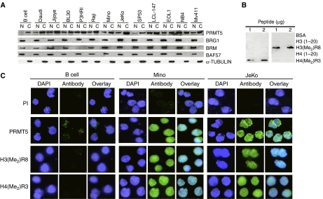
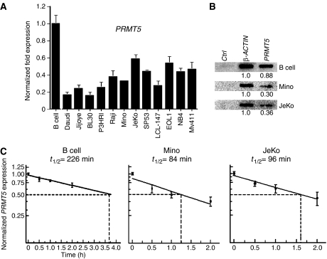
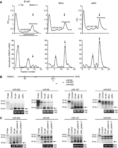
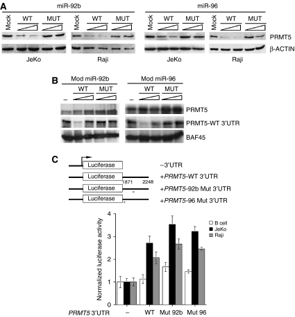
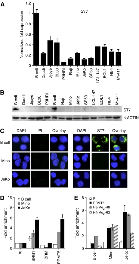
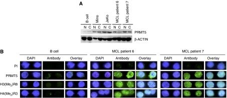
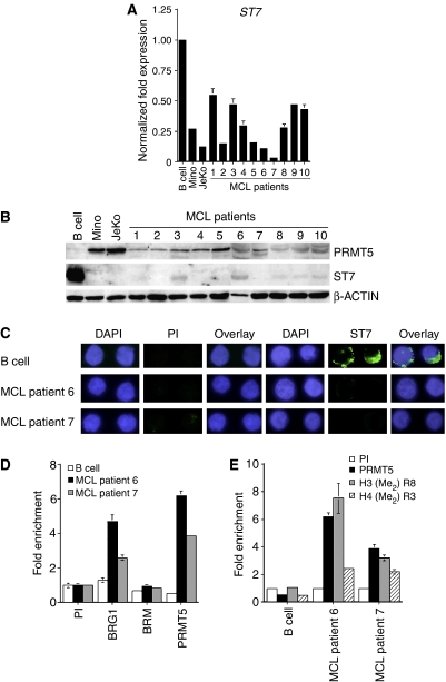
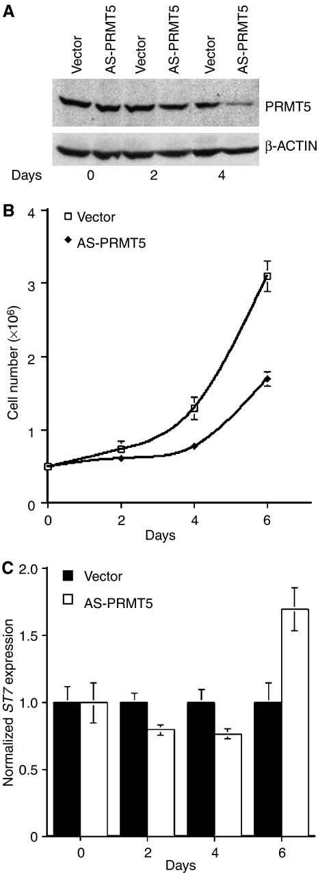
Protein arginine methyltransferase PRMT5 interacts with human SWI/SNF complexes and methylates histones H3R8 and H4R3. To elucidate the role of PRMT5 in human cancer, we analyzed PRMT5 expression in normal human B lymphocytes and a panel of lymphoid cancer cell lines as well as mantle cell lymphoma (MCL) clinical samples. We show that PRMT5 protein levels are elevated in all cancer cells, including clinical samples examined despite its low rate of transcription and messenger RNA stability. Remarkably, polysome profiling revealed that PRMT5 mRNA is translated more efficiently in Mino and JeKo MCL cells than in normal B cells, and that decreased miR-92b and miR-96 expression augments PRMT5 translation. Consequently, global methylation of H3R8 and H4R3 is increased and is accompanied by repression of suppressor of tumorigenecity 7 (ST7) in lymphoid cancer cells. Furthermore, knockdown of PRMT5 expression reduces proliferation of transformed JeKo and Raji cells. Thus, our studies indicate that aberrant expression of PRMT5 leads to altered epigenetic modification of chromatin, which in turn impacts transcriptional performance of anti-cancer genes and growth of transformed lymphoid cells.


Figures

Figure 1
Figure 1
PRMT5 is overexpressed in lymphoma and leukemia cell lines. (A) Expression of PRMT5 and hSWI/SNF subunits were assessed by Western blotting using either nuclear (N) and cytosolic (C) extracts from normal CD19+ B cells, or the indicated transformed cell lines (20 μg). To discern between N and C fractions, we measured α-TUBULIN expression. (B) Anti-H3(Me2)R8 and anti-H4(Me2)R3 antibodies do not crossreact and are highly specific. Portions (1 and 2 μg) of BSA or the indicated peptides were spotted on nitrocellulose membrane, and detected using anti-H4(Me2)R3 or anti-H3(Me2)R8. (C) Immunofluorescence of PRMT5, H4(Me2)R3, and H3(Me2)R8 in normal B cells, Mino, and JeKo cells. Normal B and MCL cells were fixed and incubated with either pre-immune, or immune anti-PRMT5, anti-H3(Me2)R8, and H4(Me2)R3 antibodies. FITC-labeled goat anti-rabbit antibody was used to detect PRMT5 and modified histones, and DAPI was used to stain nuclei. Pictures were taken at × 100 magnification.
Figure 2
Figure 2
Expression of PRMT5 is regulated at different levels. (A) PRMT5 mRNA expression was measured by real-time RT–PCR in normal as well as transformed B cells. The bar graph shows normalized fold expression of PRMT5 mRNA in various cell lines relative to normal B cells using GAPDH as internal control. RT–PCR analysis was performed three times in triplicates and the graph shows the average±s.d. (B) The rate of PRMT5 transcription is altered in MCL cell lines. Nuclear run on assays were conducted as described in materials and methods. Control (Ctrl) PvuII–PvuII DNA fragment of pBluescript KS(+), and β-ACTIN and PRMT5 cDNA PCR fragments were immobilized on Hybond XL membrane and detected with radiolabeled RNA isolated from the indicated cells. Signals were quantitated and expression of PRMT5 was reported relative to β-ACTIN. (C) PRMT5 mRNA is more stable in B cells. Normal and transformed B cells were treated with DRB for the indicated times, and total RNA was isolated and quantitated by real-time RT–PCR. PRMT5 mRNA expression was normalized using 18S as internal control. To calculate the half-life (t1/2) of PRMT5 mRNA, data points in each graph were generated from four distinct experiments conducted in triplicates.
Figure 3
Figure 3
PRMT5 mRNA is translated more efficiently in MCL cell lines. (A) Polyribosome profiles of normal and transformed B cells. Whole-cell lysate were fractionated by 15–40% sucrose gradient sedimentation, and polyribosome profiles were determined by measuring the absorbance of each fraction at 254 nm (upper panel). Fractions representing 40S, 60S, 80S and polyribosomes are indicated. RNA isolated from each fraction was used to measure the level of PRMT5 mRNA by real-time RT–PCR, and β-ACTIN was used as an internal control to normalize PRMT5 mRNA levels in each fraction (lower panel). The amount of PRMT5 mRNA present in each fraction is reported relative to the fraction containing the lowest copy number of PRMT5 mRNA. The data points in each graph represent the average from triplicate RT–PCR reactions±s.d. Vertical arrows indicate peak polyribosome fractions. (B, C) Differential expression of PRMT5-specific miRNAs in normal and transformed B cells. Schematic representation of PRMT5 mRNA depicting the position of potential miRNA-binding sites within the 3′UTR (B, upper panel). RPA was performed on 20 μg (B) or 5 μg (C) of total RNA isolated from the indicated cells using miR-92b, miR-96, miR-197, or miR-607 probe. Probe represents 1/10th of the total amount of labeled probe used in each reaction, and control shows digestion of the probe in presence of yeast tRNA. Ethidium bromide-stained gels are included to show equal loading. Arrows show position of mature miRNAs.
Figure 4
Figure 4
PRMT5 expression is downregulated by miR-92b and miR-96 in transformed B cells. (A) JeKo and Raji cells were electroporated with 2.5 and 5.0 μg of either wild-type or mutant miR-92b and miR-96 double-stranded RNA individually, and 20 μg of RIPA extracts were analyzed by Western blotting. (B) Effect of modified miR-92b and miR-96 on PRMT5 expression in vitro. In vitro translation was carried out in the absence or presence of increasing amounts of modified wild-type or mutant miR-92b or miR-96 using 0.25 μg of PRMT5 mRNA without 3′UTR (PRMT5) and with wild-type 3′UTR (PRMT5-WT 3′UTR). BAF45 mRNA was used as a control. (C) Normal B (25 × 106) or transformed JeKo and Raji (5 × 10⁶) cells were electroporated in the presence of pRL-TK with either pCMV-LUC, pCMV-LUC fused to wild-type PRMT5 3′UTR, or pCMV-LUC fused to mutated miR-92b or miR-96-binding site PRMT5 3′UTR construct, and luciferase expression was measured using dual luciferase reporter assay. Luciferase activity is represented relative to pCMV-LUC for each cell line, and has been normalized using Renilla luciferase.
Figure 5
Figure 5
ST7 is silenced in transformed lymphoid cell lines. (A) ST7 transcription is repressed in lymphoid cancer cell lines as determined by real-time RT–PCR. This experiment has been repeated three times in triplicates. (B) ST7 protein expression was analyzed by Western blotting using 20 μg of RIPA extracts from either normal B cells, or the indicated transformed lymphoid cell lines. β-ACTIN levels were detected to ensure equal loading. (C) Fixed normal or transformed B cells were incubated with either pre-immune or immune anti-ST7 antibody. ST7 protein was visualized using goat FITC-labeled anti-rabbit antibody, whereas nuclei were stained with DAPI. Pictures were taken at × 100 magnification. (D, E) ChIP assays were performed on crosslinked chromatin from normal or transformed B cells using either preimmune (PI) or the indicated immune antibodies, and the retained DNA was amplified by real-time PCR using ST7-specific primers and probe. Fold enrichment with each antibody was calculated relative to the PI sample. Each ChIP experiment was repeated twice in triplicates.
Figure 6
Figure 6
MCL patients overexpress PRMT5 protein. (A) Nuclear and cytosolic extracts from normal CD19+ B cells or MCL clinical samples 6 and 7 (20 μg) were analyzed by Western blotting using either anti-PRMT5 or control β-ACTIN antibody. (B) Immunofluorescence of normal B and transformed MCL cells from clinical samples 6 and 7 after staining with DAPI, PI, or antibodies to PRMT5, H3(Me2)R8, and H4(Me2)R3.
Figure 7
Figure 7
Overexpression of PRMT5 correlates with ST7 silencing in MCL clinical samples. (A) Real-time RT–PCR analysis of ST7 mRNA expression in MCL patient samples 1–10. Expression of ST7 was normalized using GAPDH as an internal control. (B) Western blot analysis was performed on 20 μg of RIPA extracts from normal B cells, Mino, JeKo, and MCL clinical samples 1–10 using the indicated antibodies. (C) Immunofluorescence of normal B cells and MCL clinical samples 6 and 7 cells after staining with DAPI, PI, or immune anti-ST7 antibody. Pictures were taken at × 100 magnification. (D, E) ChIP was performed on crosslinked chromatin from normal B cells and MCL clinical samples 6 and 7 using either PI or the indicated immune antibodies. Immunoprecipitated DNA was amplified by real-time PCR, and the fold enrichment with each antibody was calculated relative to the PI sample.
Figure 8
Figure 8
Knocking down PRMT5 expression affects growth of transformed B cells. (A) Western blot analysis was performed on 20 μg of RIPA extract from JeKo cells after infection with either vector or AS-PRMT5 lentivirus for 0, 2, and 4 days using anti-PRMT5 and control anti-β-ACTIN antibodies. (B) Proliferation of JeKo cells infected with either control vector or AS-PRMT5 lentivirus. Cells were counted every two days for 6 days, and the experiment was repeated four times in duplicates. (C) ST7 mRNA expression in lentivirus infected JeKo cells was evaluated by real-time RT–PCR at the indicated times. ST7 mRNA expression is represented relative to control vector infected JeKo cells, and is normalized to GAPDH.

References

    1. Ancelin K, Lange UC, Hajkova P, Schneider R, Bannister AJ, Kouzaride T, Surani MA (2006) Blimp1 associates with Prmt5 and directs histone arginine methylation in mouse germ cells. Nat Cell Biol 8: 623–630 - PubMed
    1. Bedford MT, Richard S (2005) Arginine methylation an emerging regulator of protein function. Mol Cell 18: 263–272 - PubMed
    1. Cao R, Wang L, Wang H, Xia L, Erdjument-Bromage H, Tempst P, Jones RS, Zhang Y (2002) Role of histone H3 lysine 27 methylation in polycomb -group silencing. Science 298: 1039–1043 - PubMed
    1. Dacwag CS, Ohkawa Y, Pal S, Sif S, Imbalzano AN (2007) The protein arginine methyltransferase Prmt5 is required for myogenesis because it facilitates ATP-dependent chromatin remodeling. Mol Cell Biol 27: 384–394 - PMC - PubMed
    1. Esteller M (2006) Epigenetics provides a new generation of oncogenes and tumour-suppressor genes. Br J Cancer 94: 179–183 - PMC - PubMed

Publication types

LinkOut - more resources