. 2007 Aug;117(8):2145-54.
doi: 10.1172/JCI31752.

Enhanced at puberty 1 (EAP1) is a new transcriptional regulator of the female neuroendocrine reproductive axis

Affiliations

Enhanced at puberty 1 (EAP1) is a new transcriptional regulator of the female neuroendocrine reproductive axis

Sabine Heger et al. J Clin Invest. 2007 Aug.

Abstract

The initiation of mammalian puberty and the maintenance of female reproductive cycles are events controlled by hypothalamic neurons that secrete the decapeptide gonadotropin-releasing hormone (GnRH). GnRH secretion is, in turn, controlled by changes in neuronal and glial inputs to GnRH-producing neurons. The hierarchical control of the process is unknown, but it requires coordinated regulation of these cell-cell interactions. Here we report the functional characterization of a gene (termed enhanced at puberty 1 [EAP1]) that appears to act as an upstream transcriptional regulator of neuronal networks controlling female reproductive function. EAP1 expression increased selectively at puberty in both the nonhuman primate and rodent hypothalamus. EAP1 encoded a nuclear protein expressed in neurons involved in the inhibitory and facilitatory control of reproduction. EAP1 transactivated genes required for reproductive function, such as GNRH1, and repressed inhibitory genes, such as preproenkephalin. It contained a RING finger domain of the C3HC4 subclass required for this dual transcriptional activity. Inhibition of EAP1 expression, targeted to the rodent hypothalamus via lentivirus-mediated delivery of EAP1 siRNAs, delayed puberty, disrupted estrous cyclicity, and resulted in ovarian abnormalities. These results suggest that EAP1 is a transcriptional regulator that, acting within the neuroendocrine brain, contributes to controlling female reproductive function.


Figures

Figure 1
Figure 1. EAP1 mRNA abundance increases in the nonhuman primate and rat hypothalamus at the time of puberty.
(A) Puberty-related increase in EAP1 mRNA content in the hypothalamus of female rhesus monkeys detected using human cDNA microarrays. Changes in mRNA content are expressed as fold increase over values present in prepubertal juvenile (Juv) animals. EP, early puberty; MP, mid-puberty. (B) EAP1 mRNA abundance increases in the MBH of female rhesus monkeys at the time of puberty, as assessed by real-time PCR. (C) EAP1 mRNA content does not increase in the CTX. ***P < 0.01 versus Juv control. (D) Abundance of the mRNA encoding the EAP1 homolog CAB45750 does not change in the MBH at the time of puberty. EAP1 mRNA content increases in the MBH (E), but not in the CTX (F), of female rats at the time of puberty, as assessed by semiquantitative PCR. Numbers above bars represent number of animals per group, and error bars are SEM. **P < 0.02 versus Juv control. Filled circles in A represent the range of values reported in the 2 microarrays and that were subsequently used to compare EP and MP animals with the Juv group; open circles in panel C represent the individual values of the only 2 animals used in this group.
Figure 2
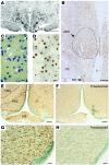
Figure 2. The EAP1 gene is expressed in subregions of the nonhuman primate and rat hypothalamus involved in reproductive control.
(A) Low-magnification image showing that EAP1 mRNA expression is prominent in cells of the ARC of the primate hypothalamus as assessed by in situ hybridization using a monkey-specific [35S]UTP-labeled EAP1 cRNA probe. The section shown is from an 8-year-old adult female monkey. ME, median eminence of the hypothalamus; OC, optic chiasm; V, third ventricle. Scale bar: 300 μm. (B) Montage image of the monkey hypothalamus (adult, 8-year-old female monkey) showing that EAP1 immunoreactive protein is preferentially localized to cells of the ARC. The dotted line outlines the medial and lateral ARC. Scale bar: 200 μm. (C) High-magnification view of A showing that EAP1 mRNA is localized to cell nuclei that appear to be neuronal because they are lightly stained with thionin (examples denoted by arrows). Scale bar: 20 μm. (D) High-magnification view of the ARC showing immunoreactive EAP1 in neuronal nuclei. Scale bar: 20 μm. (E) Low-magnification view of the MBH from a 28-day-old prepubertal female rat showing EAP1 staining in cell nuclei of the ARC and ventromedial (VMH) nuclei (outlined by dotted lines). The immunohistochemical reaction was developed to a brown color with diaminobenzidine hydrochloride (Supplemental Note 3), and the cell nuclei were counterstained with 0.1% methyl green. (F) Lack of EAP1 staining in an adjacent section incubated with EAP1 antibodies preadsorbed with the antigenic peptide used to prepare the antibodies (10 μg peptide/ml antiserum, overnight at 4°C). (G) Higher-magnification view of the ARC showing EAP1 staining (brown) in cell nuclei. Some cells did not contain EAP1, so their nuclei have only a green color. (H) Adjacent section stained with peptide-preadsorbed EAP1 antibodies. Scale bars: 100 μm (in E and F); 20 μm (G and H).
Figure 3
Figure 3. EAP1 is a nuclear protein with dual transcriptional activity expressed in hypothalamic neurons that facilitate or inhibit the initiation of female puberty.
(A) Confocal image of a subregion of the rat POA from a 28-day-old female rat showing that EAP1 has a predominant nuclear localization (green). Notice that not all neurons contain EAP1, as evidenced by the lack of green staining in cell nuclei identified by Hoechst staining (blue). Long arrows indicate EAP1-positive cells; short arrows indicate cells lacking EAP1. (B) Detection of EAP1 (green) in the nucleus of GnRH neurons (red, arrows). (C) Wide-field image of a neuron labeled for EAP1 and Hoechst after constrained iterative deconvolution and 3D reconstruction showing that EAP1 (red) is not associated with condensed chromatin (blue). Scale bars: 40 μm (A); 20 μm (B); 5 μm (C). (DF) Combined immunohistochemistry–in situ hybridization of brain sections from 28- to 30-day-old female rats showing that EAP1 protein is abundant (D) in cells of the latero-ventral portion of the ventromedial nucleus (LVMH) of the hypothalamus, identified as enkephalinergic by their content of preproenkephalin (preproEnk) mRNA (E and F). The higher-magnification image in F shows that all preproenkephalin mRNA–containing neurons (white grains) are EAP1 positive (brown staining; examples denoted by arrows). 3V, third ventricle. (GI) EAP1 has a dual transcriptional regulatory activity that requires an intact RING finger domain. (G) In GT1-7 GnRH-producing cells, EAP1 transactivates the GnRH promoter. (H) In the same cells, it represses the preproenkephalin promoter. (I) This repressive activity is also seen in hippocampal neuroprogenitor HiB5 cells. Both the transactivating and repressive activities of EAP1 are abolished by either a single amino acid substitution (C→A) in the EAP1 RING finger domain or by deletion of this domain (ΔR). Numbers above bars represent number of wells/group, and error bars are SEM.
Figure 4
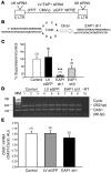
Figure 4. Lentivirus-mediated delivery of EAP1 siRNAs decreases EAP1 mRNA expression both in vitro and in vivo.
(A) Diagram of the lentivirus construct (27) used, showing the site of insertion of a U6 promoter–driven shRNA-expressing cassette. (B) Generation of EAP1 sh1 from the U6 promoter–directed transcriptional cassette. (C) Inhibition of EAP1 gene expression in HiB5 hippocampal neural progenitor cells by EAP1 sh1 and -3, measured 48 hours after infection. EAP1 mRNA content was detected by semiquantitative PCR. Bars are mean ± SEM; n = 6–7 wells/group, except in the case of noninfected control (n = 3). *P < 0.05; ***P < 0.01 versus control or LV-infected group. (D and E) Lack of changes in 2ι,5ι oligoadenylate synthetase–1 (OAS1) mRNA abundance after in vitro infection of hypothalamic slices with lentiviral particles carrying EAP1 sh1. The slices were prepared and infected as described in Supplemental Note 7. OAS1 mRNA was measured by semiquantitative PCR 72 hours after infection. (D) PCR products size-fractionated in a 2% agarose gel stained with ethidium bromide. –RT, no reverse transcription product. MM, molecular markers. (E) Densitometric analysis of the gel shown in D. Cyclophilin (Cyclo) mRNA, a housekeeping gene, was used as the normalizing unit. Accordingly, OAS1 mRNA levels are expressed as mean ± SEM (AU) of the OAS1/cyclophilin mRNA ratio calculated for each group (n = 3).
Figure 5
Figure 5. In vivo lentivirus-mediated delivery of EAP1 shRNAs to the rat POA decreases EAP1 protein levels.
The images illustrate results derived from 8 control animals injected with LV eGFP, 6 animals correctly injected with EAP1 sh1, and 2 animals with misplaced injections of EAP1 sh1. (A and B) Examples of well-placed injections carrying EAP1 sh1–encoding lentiviruses. The cells infected are seen along the lateral edge (A) or dorsal-medial aspect (B) of the AVPV. (C) Example of misplaced injections (too lateral and too caudal). (D) Higher-magnification image showing that most infected cells had neuronal morphology. (E) Example of an infected GnRH neuron (yellow). (FI) EAP1 protein content (red) was reduced in neurons infected with EAP1 sh1 (green). (F) The injection track, rich in eGFP-expressing neurons, shows fewer EAP1-positive cells (red) than neighboring areas. (G) Higher-magnification images illustrating the reduction in EAP1 immunoreactive material (red) in the nucleus of EAP1 sh1–infected cells (eGFP-positive, green) as compared with noninfected (eGFP-negative, red only) cells. (H) Same field as in G, but showing only EAP1 immunoreactivity (red) to illustrate the loss of EAP1 protein in EAP1 sh1–infected cells. (I) Fluorescence intensities in AUs based on measurements of 36 infected and 104 noninfected cells. ***P < 0.01 versus noninfected cells. Scale bars: 20 μm (D); 5 μm (E); 50 μm (F); 10 μm (G and H). Numbers in parentheses represent number of wells per group, and error bars are SEM. Arrows in AC point to sites of injection; arrows in G and H point to infected cells (G) exhibiting a decrease in EAP1 immunoreactivety (H).
Figure 6
Figure 6. Site-restricted shRNA-mediated knockdown of the EAP1 gene targeted to the rat POA delays puberty, disrupts estrous cyclicity, and reduces plasma gonadotropin and estradiol levels.
EAP1-knockdown animals exhibit: delayed puberty, assessed by the age of first ovulation (A); disruption of estrous cyclicity, determined in daily vaginal lavages (B and C, example of a disrupted cycle); episodes of persistent estrus (D); decreased serum LH (E), FSH (F), and estradiol levels (G). Numbers above bars represent number of animals per group, and error bars are SEM. *P < 0.05, **P < 0.02, and ***P < 0.01 versus LV eGFP–injected controls.
Figure 7
Figure 7. Site-restricted shRNA-mediated knockdown of the EAP1 gene targeted to the rat POA results in abnormal follicular development.
(A) Normal ovarian follicle growth is disrupted as evidenced by an accumulation of antral follicles. (B) The accumulation is due to an increased number of medium-size antral follicles in the 200- to 400-μm range. (C) Example of an ovary from an LV eGFP–injected control rat showing a normal complement of antral follicles (AF) and corpora lutea (CL). (D) The ovaries from EAP1 sh1–injected rats exhibited follicular and luteal cysts (FC and LC, respectively). (E) Higher-magnification view of D. Each bar represents mean ± SEM of 3 animals. *P < 0.05 versus LV eGFP–injected controls.

Similar articles

Cited by

References

    1. Plant, T.M., and Witchel, S.F. 2006. Puberty in nonhuman primates and humans. In The physiology of reproduction. 3rd edition. J.D. Neill, editor. Academic Press/Elsevier. San Diego, California, USA. 2177–2230.
    1. Ojeda, S.R., and Skinner, M.K. 2006. Puberty in the rat. In The physiology of reproduction. 3rd edition. J.D. Neill, editor. Academic Press/Elsevier. San Diego, California, USA. 2061–2126.
    1. Kordon, C., Drouva, S.V., Martínez de la Escalera, G., and Weiner, R.I. 1994. Role of classic and peptide neuromediators in the neuroendocrine regulation of luteinizing hormone and prolactin. In The physiology of reproduction. 2nd edition. Volume 1. E. Knobil and J.D. Neill, editors. Raven Press. New York, New York, USA. 1621–1681.
    1. Ojeda, S.R., and Terasawa, E. 2002. Neuroendocrine regulation of puberty. In Hormones, brain and behavior. Volume 4. D. Pfaff et al., editors. Elsevier. New York, New York, USA. 589–659.
    1. Ojeda S.R., et al. Glia-to-neuron signaling and the neuroendocrine control of female puberty. Ann. Med. 2003;35:244–255. - PubMed

Publication types

MeSH terms