. 2007 Jul;5(4):394-402.
doi: 10.2174/157016207781024018.

Rev-dependent indicator T cell line

Affiliations

Rev-dependent indicator T cell line

Yuntao Wu et al. Curr HIV Res. 2007 Jul.

Abstract

Measuring virion infectivity is critical for studying and monitoring the process of HIV-1 infection. The easiest and the most common method utilizes reporter cell lines based on the HIV LTR promoter. The early HIV gene product Tat amplifies expression from the LTR; however, there is a background transcriptional activity that is independent of Tat. Furthermore, LTR activity can be influenced by cellular activation states. We have recently constructed a Rev-dependent expression vector, and as a test of this construct's functionality, we have integrated this vector into a continuous T cell line. This novel indicator cell has no measurable background signal, is not affected by elevated metabolic states, and yet responds robustly to the presence of HIV. The line is able to complete TCID50 assays in 3-5 days, and appears sensitive to both CCR5- and CXCR4-utilizing viruses.


Figures

Fig (1)
Fig (1)
Rev-dependent reporter construct. (A) The vector includes four separate segments of the HIV genome with no intact HIV genes. The 5′ end of the vector consists of the HIV 5′ LTR, the splice donor 1 site, D1, and a portion of the gag open reading frame that includes the packaging signal. The second HIV segment is from the tat1/rev1 exon that includes splice acceptor site 5, A5, and splice donor site 4, D4. The third segment of HIV DNA is from the env exon and encompasses the RRE, and the splice acceptor site 7, A7. The last segment includes the entire 3′ LTR along with a small portion of the nef reading frame 5′ to the LTR. In the absence of HIV infection the reporter provirus undergoes basal transcription generating a single message that is rapidly spliced, and results in the removal of the GFP reading frame. In the presence of HIV Rev, singly and non-spliced transcript are delivered to the cytosol, and the reporter gene is expressed. (B) CEM-SS cells were infected with the Rev-dependent green fluorescent protein (GFP) reporter construct incorporated into a lentivirus. A clone was isolated and infection by HIV resulted in generation of GFP.
Fig (2)
Fig (2)
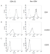
Flow cytometry analyses of Rev-CEM cells for surface expression of HIV-1 receptor and co-receptors. Rev-CEM and control CEM-SS cells were stained with R-phycoerythrin (A,C) or FITC (B) conjugated monoclonal antibodies (solid lines) against CD4 (A), CXCR4 (B) or CCR5 (C). The isotype antibody staining of the cells is shown in dotted lines. The isotype and CCR5 cytometry analysis of the CEM-SS cells overlapped so that the two runs are not discernable. Twenty thousand cells were examined in each run. Only viable cells are displayed. Two independent analyses yielded the same result.
Fig (3)
Fig (3)
HIV-independent induction of GFP expression in LTR-based CEM-GFP and Rev-CEM cells. To examine non-specific induction of the reporter CEM-GFP (blue lines) or Rev-CEM (red lines) cells were stimulated (solid line) or not (dashed line) with PMA (100ng/ml) for 24 hrs, then analyzed by flow cytometry for GFP expression. Twenty thousand cells were examined in each run.
Fig (4)
Fig (4)
Comparison of reporter generation by CEM-GFP and Rev-CEM cells following low level HIV infection. The LTR-based CEM-GFP (A and B) and Rev-CEM (C and D) cell populations were either not infected (A and C) or were infected (B and D) with equivalent levels (see Methods) of HIVNL4-3. After 3 days the cells were fixed and fluorescence from GFP (FL1) was determined. Gates were positioned to minimize background events from the non-HIV control populations (less than 0.1%). The fold-increase in fluorescence intensity (geometric mean) of non-infected cells (A and C) compared to the gated populations of GFP-positive cells in HIV-infected cells (B and D) was determined for both the CEM-GFP (n=2, 30.6- and 35.7-fold) and the Rev-CEM line (n=4, 11.6 ± 1.5; mean ± SE).
Fig (5)
Fig (5)
Transcriptional analyses of Rev-CEM in response to HIV-1 infection. (A) diagram of PCR primers used. Two sets of primers were used to detect the spliced (black arrows) and unspliced (green arrows) transcripts by reverse transcriptase PCR (RT-PCR) as described in Methods. The mRNA molecules from HIV-infected (82 ng p24; HIVNL4-3/0.5 million cells) and uninfected cells were extracted and analyzed by RT-PCR for the spliced transcripts and unspliced transcripts (B). CEM-SS cells were used as a control for the specificity of PCR amplification. The cellular β-actin transcript was also amplified to ensure that comparable numbers of cells were being used for the analyses.

References

    1. Aguilarcordova E, Chinen J, Donehower L, Lewis DE, Belmont JW. A sensitive reporter cell-line for HIV-1 Tat activity, HIV-1 inhibitors, and T-cell activation effects. Aids Research and Human Retroviruses. 1994;10:295–301. - PubMed
    1. Czerniecki BJ, Carter C, Rivoltini L, Koski GK, Kim HI, Weng DE, Roros JG, Hijazi YM, Xu SW, Rosenberg SA, Cohen PA. Calcium ionophore-treated peripheral blood monocytes and dendritic cells rapidly display characteristics of activated dendritic cells. Journal of Immunology. 1997;159:3823–3837. - PubMed
    1. Dorsky DI, Wells M, Harrington RD. Detection of HIV-1 infection with a green fluorescent protein reporter system. Journal of Acquired Immune Deficiency Syndromes and Human Retrovirology. 1996;13:308–313. - PubMed
    1. Englund G, Theodore TS, Freed EO, Engelman A, Martin MA. Integrationis required for productive infection of monocyte-derived macrophages by human-immunodeficiency-virus type-1. Journal of Virology. 1995;69:3216–3219. - PMC - PubMed
    1. Felber BK, Pavlakis GN. A quantitative bioassay for HIV-1 based on trans-activation. Science. 1988;239:184–187. - PubMed

Publication types

Substances