

Age-related changes in the occurrence and characteristics of thymic CD4(+) CD25(+) T cells in mice
- PMID: 17627771
- PMCID: PMC2266020
- DOI: 10.1111/j.1365-2567.2007.02667.x
Age-related changes in the occurrence and characteristics of thymic CD4(+) CD25(+) T cells in mice
Abstract
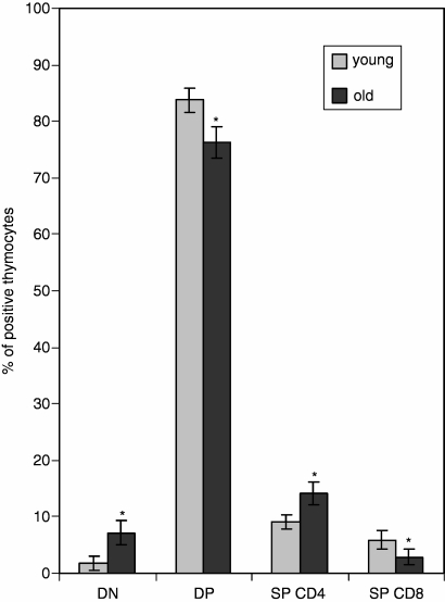
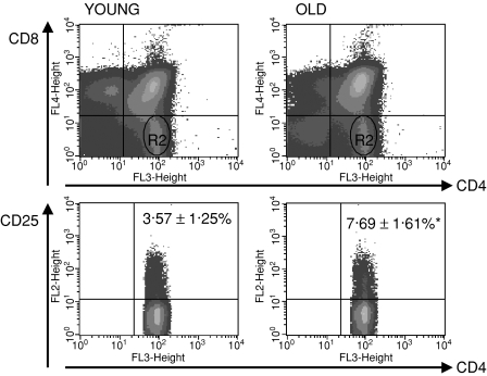
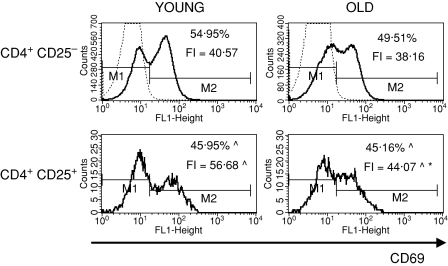
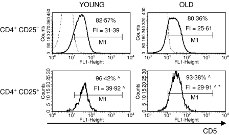
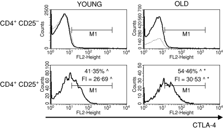
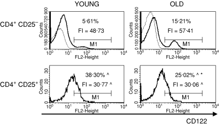
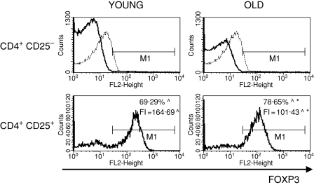
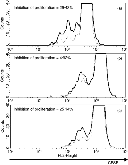
Natural regulatory CD4(+) CD25(+) T cells play an important role in preventing autoimmunity by maintaining self-tolerance. They express CD25 constitutively and are produced in the thymus as a functionally mature T-cell population. Changes in the potential of these cells to regulate the activity of conventional effector lymphocytes may contribute to an increased susceptibility to infection, cancer and age-associated autoimmune diseases. In this study we demonstrated that the thymi of aged mice are populated by a higher percentage of CD4(+) CD25(+) thymocytes than in young animals. The expression of several surface markers (CD69, CD5, CD28, CTLA-4, CD122, FOXP3), usually used to characterize the phenotype of CD4(+) CD25(+) T regulatory cells, was compared between young and aged mice. We also examined the ability of sorted thymus-deriving regulatory T cells of young and aged BALB/c mice to inhibit the proliferation of lymph node lymphocytes activated in vitro. Natural regulatory T cells isolated from the thymi of young mice suppress the proliferation of responder lymph node cells. We demonstrated that thymus-deriving CD4(+) CD25(+) T cells of old mice maintain their potential to suppress the proliferation of activated responder lymphocytes of young mice. However, their potential to inhibit the proliferation of old responder T cells is abrogated. Differences in the occurrence and activity of CD4(+) CD25(+) thymocytes between young and old animals are discussed in relation to the expression of these surface markers.
Figures

Similar articles
-
Thymus and autoimmunity: production of CD25+CD4+ naturally anergic and suppressive T cells as a key function of the thymus in maintaining immunologic self-tolerance.J Immunol. 1999 May 1;162(9):5317-26. J Immunol. 1999. PMID: 10228007
-
The significantly enhanced frequency of functional CD4+CD25+Foxp3+ T regulatory cells in therapeutic dose aspirin-treated mice.Transpl Immunol. 2009 Mar;20(4):253-60. doi: 10.1016/j.trim.2008.12.001. Epub 2009 Jan 13. Transpl Immunol. 2009. PMID: 19146957
-
Development of mouse CD4(+)CD25(+)Foxp3(+) regulatory T cells in xenogeneic pig thymic grafts.Transpl Immunol. 2009 Jan;20(3):180-5. doi: 10.1016/j.trim.2008.09.006. Epub 2008 Oct 7. Transpl Immunol. 2009. PMID: 18845256
-
Thymic regulatory T cells.Autoimmun Rev. 2005 Nov;4(8):579-86. doi: 10.1016/j.autrev.2005.04.010. Autoimmun Rev. 2005. PMID: 16214099 Review.
-
Induction and maintenance of self tolerance: the role of CD4+CD25+ regulatory T cells.Arch Immunol Ther Exp (Warsz). 2006 Sep-Oct;54(5):307-21. doi: 10.1007/s00005-006-0035-x. Epub 2006 Oct 6. Arch Immunol Ther Exp (Warsz). 2006. PMID: 17031470 Review.
Cited by
-
Mitigating age-related immune dysfunction heightens the efficacy of tumor immunotherapy in aged mice.Cancer Res. 2012 Apr 15;72(8):2089-99. doi: 10.1158/0008-5472.CAN-11-3019. Epub 2012 Apr 11. Cancer Res. 2012. PMID: 22496463 Free PMC article.
-
Prolonged graft survival in older recipient mice is determined by impaired effector T-cell but intact regulatory T-cell responses.PLoS One. 2010 Feb 16;5(2):e9232. doi: 10.1371/journal.pone.0009232. PLoS One. 2010. PMID: 20169060 Free PMC article.
-
Age effects of distinct immune checkpoint blockade treatments in a mouse melanoma model.Exp Gerontol. 2018 May;105:146-154. doi: 10.1016/j.exger.2017.12.025. Epub 2018 Jan 8. Exp Gerontol. 2018. PMID: 29326088 Free PMC article.
-
Critical influence of the thymus on peripheral T cell homeostasis.Immun Inflamm Dis. 2016 Nov 28;4(4):474-486. doi: 10.1002/iid3.132. eCollection 2016 Dec. Immun Inflamm Dis. 2016. PMID: 27980781 Free PMC article.
-
Immunosenescence, Physical Exercise, and their Implications in Tumor Immunity and Immunotherapy.Int J Biol Sci. 2025 Jan 6;21(3):910-939. doi: 10.7150/ijbs.100948. eCollection 2025. Int J Biol Sci. 2025. PMID: 39897036 Free PMC article. Review.
References
-
- Sakaguchi S, Sakaguchi N, Asano M, et al. Immunologic self tolerance maintained by activated T cells expressing IL-2 receptor α-chains (CD25): breakdown of a single mechanism of self-tolerance causes various autoimmune diseases. J Immunol. 1995;160:1151–64. - PubMed
-
- Itoh M, Takahashi T, Sakaguchi N, et al. Thymus and autoimmunity: production of CD25+CD4+ naturally anergic and suppressive T cells as a key function of the thymus in maintaining immunologic self-tolerance. J Immunol. 1999;162:5317–26. - PubMed
-
- Shevach EM. Regulatory T cells in autoimmunity. Annu Rev Immunol. 2000;18:423–49. - PubMed
-
- Sakaguchi S. Regulatory T cells: key controllers of immunologic self-tolerance. Cell. 2000;101:455–8. - PubMed
-
- Fehervari Z, Sakaguchi S. Development and function of CD4+CD25+ regulatory T cells. Curr Op Immunol. 2004;16:203–8. - PubMed
Publication types
MeSH terms
Substances
LinkOut - more resources
Full Text Sources
Medical
Research Materials
