. 2007 Sep 1;583(Pt 2):731-42.
doi: 10.1113/jphysiol.2007.139352. Epub 2007 Jul 12.

Histamine excites neurones in the human submucous plexus through activation of H1, H2, H3 and H4 receptors

Affiliations

Histamine excites neurones in the human submucous plexus through activation of H1, H2, H3 and H4 receptors

Eva Breunig et al. J Physiol. .

Abstract

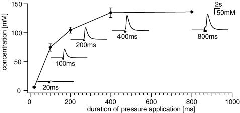
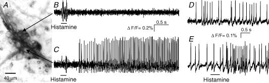
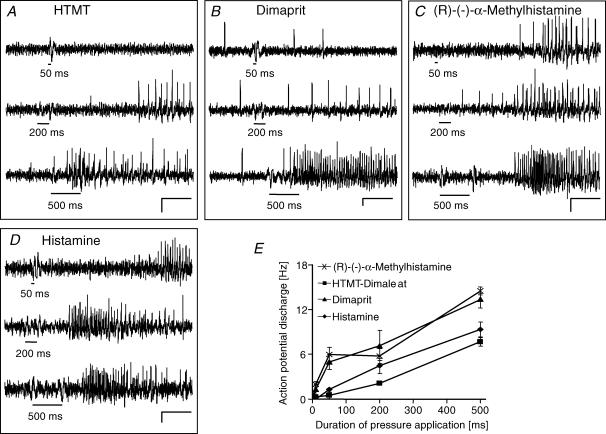
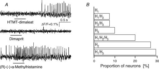
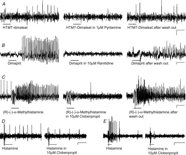
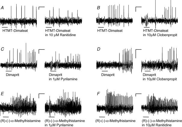
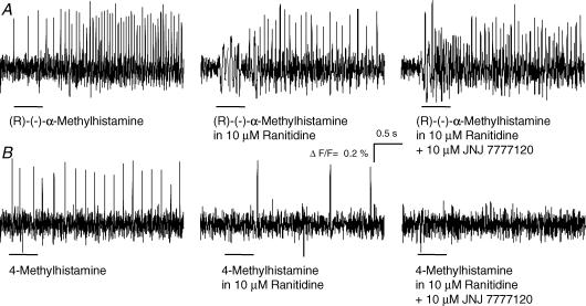
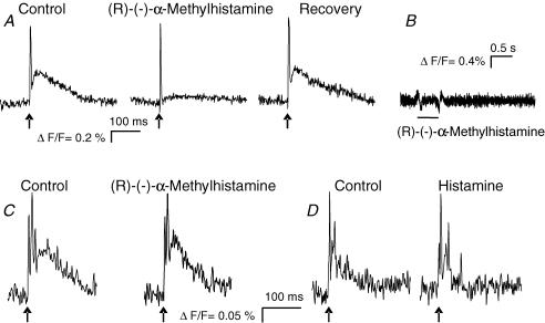
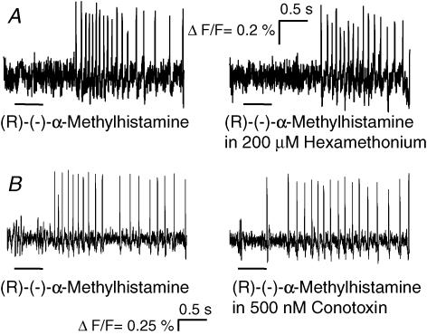
Histamine is a major mast cell mediator of immunoneural signalling in the gut and mast cells play a role in the pathophysiology of functional and inflammatory bowel diseases. Histamine receptors are therefore promising drug targets to treat gut disorders. We aimed to study the so far unknown effect of histamine on neural activity in the human enteric nervous system (ENS) and to identify the pharmacology of histamine response. We used fast imaging techniques in combination with the potentiometric dye di-8-ANEPPS to monitor directly membrane potential changes and thereby neuronal excitability in the human submucous plexus from surgical specimens of 110 patients (2137 neurones, 273 ganglia). Local microejection of histamine resulted in action potential discharge in 37% of neurones. This excitatory effect was mimicked by the H(1) agonist HTMT-dimaleat, H(2) agonist dimaprit, H(3) agonist (R)-(-)-alpha-methylhistamine and H(4) agonist 4-methylhistamine. The excitatory actions of the agonists were specifically and selectively blocked by the H(1), H(2), H(3) or H(4) receptor antagonists pyrilamine, ranitidine, clobenpropit or J1-[(5-chloro-1H-indol-2-yl)carbonyl]-4-methylpiperazine (JNJ 7777120), respectively. Clobenproprit reduced the excitatory response to histamine. Unlike in the guinea-pig ENS (R)-(-)-alpha-methylhistamine had no presynaptic actions in human submucous plexus. Application of agonists revealed receptor clustering which was as follows: 29% H(1)/H(3), 27% H(2), 20% H(1)/H(2)/H(3), 10% H(3), 7% H(1)/H(2) and 7% H(2)/H(3). Histamine excites human enteric neurones and this effect involves all four histamine receptors; most striking was the identification of an excitatory H(3) mediated component and the discovery of H(4) mediated neuronal excitation. These data may form the basis of identification of new targets to treat inflammatory and functional gut disorders.


Figures

Figure 1
Figure 1. Drug dilution with pressure pulse microejection measured with a concentration element
The duration of pressure ejection pulses of 1 m KCl (X-axis) is plotted against the calculated concentration of KCl (Y-axis) at the tip of the electrode. Insets illustrate representative traces of changes in KCl concentration for different pulse durations. Each data point represents 2–10 experiments. See Methods for further explanation.
Figure 2
Figure 2. Histamine evoked action potential discharge in a subset of human submucous neurones
A, fluorescence image of a Di-8-ANEPPS labelled submucous ganglion. Individual neurones of the ganglion can be distinguished by the strong staining of the outer membrane. B and C, the change in fluorescence intensity is shown on the right for two of these neurones to pressure application of 200 ms histamine (horizontal bars). The neurone shown in the upper trace did not respond to histamine while the neurone in the bottom trace responded after a short delay with a discharge of action potentials that lasted throughout the recording period of 4 s. D, the deflections in the traces during application of histamine are pressure ejection artefacts. E, one spontaneously active neurone from a different ganglion; 500 ms histamine application to the same neuron evoked increased spike discharge without changing the discharge pattern of ongoing activity. Note that underlying slow depolarizations of membrane potential are not detected by the AC-coupled photodiode system.
Figure 3
Figure 3. Histamine, the H1, H2 and H3 agonists evoked spike discharge which increased with duration of pressure application
A–D, the H1 agonist HTMT-dimaleat, the H2 agonist dimaprit, the H3 agonist (R)-(−)-α-methylhistamine and histamine evoked spike discharge which increased with pressure application of 50 ms, 200 ms and 500 ms (black bars mark duration of drug application). Scale bars indicate changes in fluorescence intensity (ΔF/F = 0.2%) and time (0.5 s). The deflections in the traces during application of the substances are pressure ejection artefacts. E, illustration of the results for histamine and the receptor agonists after pressure application of 10 ms, 50 ms, 200 ms and 500 ms (each data point from n = 11–90 neurons). Note that underlying slow depolarizations of membrane potential are not detected by the AC-coupled photodiode system.
Figure 4
Figure 4. Responses to histamine receptor agonists suggested distinct receptor clustering
A, the same submucous neurone responded to the H1 agonist HTMT-dimaleat and the H3 agonist (R)-(−)-α-methylhistamine, but not to the H2 agonist dimaprit (each 500 ms pressure application). This type of response corresponded to the code H1/H3. B, results of such experiments from 94 neurons revealed that most neurons responded to the H1 and H3 agonist but not to the H2 agonist. All possible codes occurred, except one, which is a response to the H1 agonist only. Note that underlying slow depolarizations of membrane potential are not detected by the AC-coupled photodiode system.
Figure 5
Figure 5. Responses to histamine and histamine receptor agonists were blocked by selective receptor antagonists
A, effect of the H1 agonist HTMT-dimaleat was blocked by the H1 antagonist pyrilamine and recovered after wash out of the antagonist. B, effect of the H2 agonist dimaprit was blocked by the H2 antagonist ranitidine and recovered after wash out of the antagonist. C, effect of the H3 agonist (R)-(−)-α-methylhistamine was blocked by the H3 antagonist clobenpropit and recovered after wash out of the antagonist. Histamine and histamine receptor agonists were applied by pressure application (black bars) before, during and after perfusion of the antagonists. D, histamine evoked spike discharge is decreased in the presence of clobenproprit. E, in this neurone clobenprobrit blocked the response to histamine. The deflections in the traces during application of the agonists are pressure ejection artefacts. The effects of the agonists were restored after wash out periods of 30 min to 1 h. Traces in A, B and C are from different neurones. Scale bars represent time (0.5 s) and changes in fluorescence intensity (ΔF/F = 0.1%). Note that underlying slow depolarizations of membrane potential are not detected by the AC-coupled photodiode system.
Figure 6
Figure 6. The excitatory effects of the histamine agonists on human submucous neurones were specific with no indication of cross-reactivity
A–B, the responses to pressure application of the H1 agonist HTMT-dimaleat (black bars) remained in the presence of the H2 antagonist ranitidine (A) or the H3 antagonist clobenpropit (B). C–D, likewise the responses to pressure application of the H2 agonist dimaprit (black bars) remained in the presence of the H1 antagonist pyrilamine (C) or the H3 antagonist clobenpropit (D). E–F, the responses to pressure application of the H3 agonist (R)-(−)-α-methylhistamine (black bars) remained in the presence of the H1 antagonist pyrilamine (E) or the H2 antagonist ranitidine (F). The deflections in the traces during application of the agonists are pressure ejection artefacts. Traces in A–E were from different neurones. Scale bars represent time (0.5 s) and changes in fluorescence intensity (ΔF/F = 0.1%). Note that underlying slow depolarizations of membrane potential are not detected by the AC-coupled photodiode system.
Figure 7
Figure 7. H3 and H4 receptor mediated excitation in the human submucous plexus was receptor specific
A, pressure application of the H3 agonist (R)-(−)-α-methylhistamine evoked action potential discharge (left panel) which was not changed during perfusion of the H2 antagonist ranitidine (centre panel) or the H4 antagonist 10 μm JNJ 7777120 (right panel). B, pressure application of the H4 agonist 4-methylhistamine evoked action potential discharge (left panel). The response was diminished but not abolished during perfusion with the H2 antagonist ranitidine (centre panel). Additional perfusion of the H4 antagonist JNJ 7777120 totally blocked the response to 4-methylhistamine (right panel). All traces are from the same neurone. The deflections in the traces during application of the agonists are pressure ejection artefacts. Note that underlying slow depolarizations of membrane potential are not detected by the AC-coupled photodiode system.
Figure 8
Figure 8. The H3 receptor agonist (R)-(−)-α-methylhistamine inhibited fast EPSPs in guinea-pig myenteric neurones but had no effect on fast EPSPs in human submucous neurons
A, electrical stimulation of an interganglionic fibre tract (arrows) evoked a compound action potential (steep upstroke) originating from nerve fibre passing through the ganglion (see Schemann et al. 2005) followed by a subthreshold fast EPSP in a guinea-pig myenteric neurone (left trace). Shortly after pressure application of (R)-(−)-α-methylhistamine, the fast EPSP was almost abolished (centre panel) and recovered several minutes after the application (right panel). B, pressure application of (R)-(−)-α-methylhistamine did not evoke any postsynaptic response in a guinea-pig myenteric neurone. C, electrical stimulation of an interganglionic fibre tract (arrows) in the human submucous plexus evoked fast EPSP (left panel). The fast EPSP remained unchanged after pressure application of (R)-(−)-α-methylhistamine (right panel). D, electrical stimulation of an interganglionic fibre tract (arrow) evoked fast EPSP triggering several action potentials in a different neurone of the human submucous plexus (left panel) which was unchanged after spritz application of histamine (right panel).
Figure 9
Figure 9. The H3 agonist (R)-(−)-α-methylhistamine directly excited human submucous neurones
A–B, the response to pressure application of (R)-(−)-α-methylhistamine remained after nicotinic blockade with hexamethonium (A) or blockade of synaptic transmission by conotoxin (B). The deflections in the traces during application of the agonists are pressure ejection artefacts. Note that underlying slow depolarizations of membrane potential are not detected by the AC-coupled photodiode system.

Similar articles

Cited by

References

    1. Bakker RA. Histamine H3-receptor isoforms. Inflamm Res. 2004;53:509–516. - PubMed
    1. Barbara G, Stanghellini V, de Giorgio R, Corinaldesi R. Functional gastrointestinal disorders and mast cells: implications for therapy. Neurogastroenterol Motil. 2006;18:6–17. - PubMed
    1. Barbara G, Stanghellini V, De Giorgio R, Cremon C, Cottrell GS, Santini D, Pasquinelli G, Morselli-Labate AM, Grady EF, Bunnett NW, Collins SM, Corinaldesi R. Activated mast cells in proximity to colonic nerves correlate with abdominal pain in irritable bowel syndrome. Gastroenterology. 2004;126:693–702. - PubMed
    1. Bischoff S, Crowe SE. Gastrointestinal food allergy: new insights into pathophysiology and clinical perspectives. Gastroenterology. 2005;128:1089–1113. - PubMed
    1. Celanire S, Wijtmans M, Talaga P, Leurs R, de Esch IJ. Keynote review: Histamine H3 receptor antagonists reach out for the clinic. Drug Discov Today. 2005;10:1613–1627. - PubMed

Publication types

MeSH terms

LinkOut - more resources