

Nitric oxide mediates the vagal protective effect on ventricular fibrillation via effects on action potential duration restitution in the rabbit heart
- PMID: 17627986
- PMCID: PMC2277035
- DOI: 10.1113/jphysiol.2007.138461
Nitric oxide mediates the vagal protective effect on ventricular fibrillation via effects on action potential duration restitution in the rabbit heart
Abstract
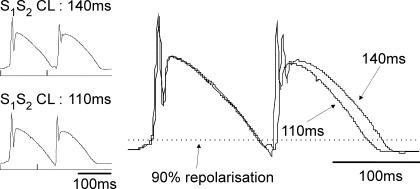
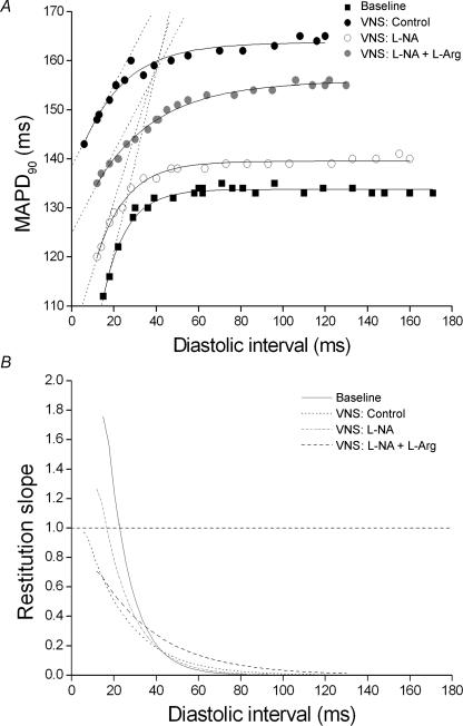
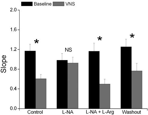
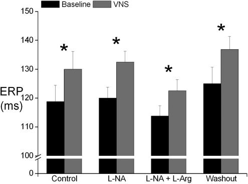
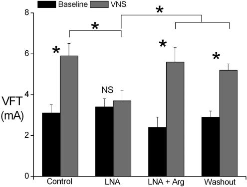
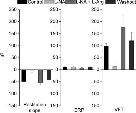
We have previously shown that direct vagus nerve stimulation (VNS) reduces the slope of action potential duration (APD) restitution while simultaneously protecting the heart against induction of ventricular fibrillation (VF) in the absence of any sympathetic activity or tone. In the current study we have examined the role of nitric oxide (NO) in the effect of VNS. Monophasic action potentials were recorded from a left ventricular epicardial site on innervated, isolated rabbit hearts (n = 7). Standard restitution, effective refractory period (ERP) and VF threshold (VFT) were measured at baseline and during VNS in the presence of the NO synthase inhibitor N(G)-nitro-L-arginine (L-NA, 200 microm) and during reversing NO blockade with L-arginine (L-Arg, 1 mm). Data represent the mean +/- S.E.M. The restitution curve was shifted upwards and became less steep with VNS when compared to baseline. L-NA blocked the effect of VNS whereas L-Arg restored the effect of VNS. The maximum slope of restitution was reduced from 1.17 +/- 0.14 to 0.60 +/- 0.09 (50 +/- 5%, P < 0.0001) during control, from 0.98 +/- 0.14 to 0.93 +/- 0.12 (2 +/- 10%, P = NS) in the presence of L-NA and from 1.16 +/- 0.17 to 0.50 +/- 0.10 (41 +/- 9%, P = 0.003) with L-Arg plus L-NA. ERP was increased by VNS in control from 119 +/- 6 ms to 130 +/- 6 ms (10 +/- 5%, P = 0.045) and this increase was not affected by L-NA (120 +/- 4 to 133 +/- 4 ms, 11 +/- 3%, P = 0.0019) or L-Arg with L-NA (114 +/- 4 to 123 +/- 4 ms, 8 +/- 2%, P = 0.006). VFT was increased from 3.0 +/- 0.3 to 5.8 +/- 0.5 mA (98 +/- 12%, P = 0.0017) in control, 3.4 +/- 0.4 to 3.8 +/- 0.5 mA (13 +/- 12%, P = 0.6) during perfusion with L-NA and 2.5 +/- 0.4 to 6.0 +/- 0.7 mA (175 +/- 50%, P = 0.0017) during perfusion with L-Arg plus L-NA. Direct VNS increased VFT and flattened the slope of APD restitution curve in this isolated rabbit heart preparation with intact autonomic nerves. These effects were blocked using L-NA and reversed by replenishing the substrate for NO production with L-Arg. This is the first study to demonstrate that NO plays an important role in the anti-fibrillatory effect of VNS on the rabbit ventricle, possibly via effects on APD restitution.
Figures
Similar articles
-
Autonomic modulation of electrical restitution, alternans and ventricular fibrillation initiation in the isolated heart.Cardiovasc Res. 2007 Mar 1;73(4):750-60. doi: 10.1016/j.cardiores.2006.12.001. Epub 2006 Dec 15. Cardiovasc Res. 2007. PMID: 17217937
-
Vagus nerve stimulation protects against ventricular fibrillation independent of muscarinic receptor activation.Cardiovasc Res. 2011 Aug 1;91(3):437-46. doi: 10.1093/cvr/cvr105. Epub 2011 May 16. Cardiovasc Res. 2011. PMID: 21576131
-
Modulation of the myocardial redox state by vagal nerve stimulation after experimental myocardial infarction.Cardiovasc Res. 2008 Mar 1;77(4):713-21. doi: 10.1093/cvr/cvm092. Epub 2007 Dec 7. Cardiovasc Res. 2008. PMID: 18065771
-
Mechanisms underlying the autonomic modulation of ventricular fibrillation initiation--tentative prophylactic properties of vagus nerve stimulation on malignant arrhythmias in heart failure.Heart Fail Rev. 2013 Jul;18(4):389-408. doi: 10.1007/s10741-012-9314-2. Heart Fail Rev. 2013. PMID: 22678767 Free PMC article. Review.
-
Vagal modulation of cardiac ventricular arrhythmia.Exp Physiol. 2014 Feb;99(2):295-9. doi: 10.1113/expphysiol.2013.072652. Epub 2013 Sep 6. Exp Physiol. 2014. PMID: 24014808 Review.
Cited by
-
Anatomical evidence of non-parasympathetic cardiac nitrergic nerve fibres in rat.J Anat. 2021 Jan;238(1):20-35. doi: 10.1111/joa.13291. Epub 2020 Aug 13. J Anat. 2021. PMID: 32790077 Free PMC article.
-
Direct evidence of nitric oxide release from neuronal nitric oxide synthase activation in the left ventricle as a result of cervical vagus nerve stimulation.J Physiol. 2009 Jun 15;587(Pt 12):3045-54. doi: 10.1113/jphysiol.2009.169417. Epub 2009 Apr 29. J Physiol. 2009. PMID: 19403619 Free PMC article.
-
QT restitution properties during exercise in male patients with coronary artery disease.Ann Noninvasive Electrocardiol. 2014 Jul;19(4):358-65. doi: 10.1111/anec.12134. Ann Noninvasive Electrocardiol. 2014. PMID: 25165790 Free PMC article.
-
Myths and realities of the cardiac vagus.J Physiol. 2013 Sep 1;591(17):4073-85. doi: 10.1113/jphysiol.2013.257758. Epub 2013 Jul 22. J Physiol. 2013. PMID: 23878363 Free PMC article. Review.
-
Ventricular arrhythmogenic remodelling in diet-induced metabolic syndrome driven by right-to-left regional differences in action potential duration and dominant frequency gradients.J Physiol. 2025 May;603(10):2979-3000. doi: 10.1113/JP286516. Epub 2025 May 5. J Physiol. 2025. PMID: 40320918 Free PMC article.
References
-
- Amlie JP, Refsum H. Vagus-induced changes in ventricular electrophysiology of the dog heart with and without beta-blockade. J Cardiovasc Pharmacol. 1981;3:1203–1210. - PubMed
-
- Bers DM. Excitation-Contraction Coupling and Cardiac Contractile Force. 1. Dordrecht: Kluwer Academic Publishers; 2001.
-
- Cao JM, Qu ZL, Kim YH, Wu TJ, Garfinkel A, Weiss JN, Karagueuzian HS, Chen PS. Spatiotemporal heterogeneity in the induction of ventricular fibrillation by rapid pacing: importance of cardiac restitution properties. Circ Res. 1999;84:1318–1331. - PubMed
-
- Chowdhary S, Townend JN. Role of nitric oxide in the regulation of cardiovascular autonomic control. Clin Sci (Lond) 1999;97:5–17. - PubMed
Publication types
MeSH terms
Substances
LinkOut - more resources
Full Text Sources
Miscellaneous