Comparative Study
. 2007 Sep 1;583(Pt 2):753-66.
doi: 10.1113/jphysiol.2007.136325. Epub 2007 Jul 12.

HIF-1alpha and HIF-2alpha play a central role in stretch-induced but not shear-stress-induced angiogenesis in rat skeletal muscle

Affiliations
Comparative Study

HIF-1alpha and HIF-2alpha play a central role in stretch-induced but not shear-stress-induced angiogenesis in rat skeletal muscle

Malgorzata Milkiewicz et al. J Physiol. .

Abstract

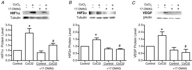
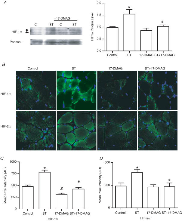
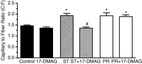
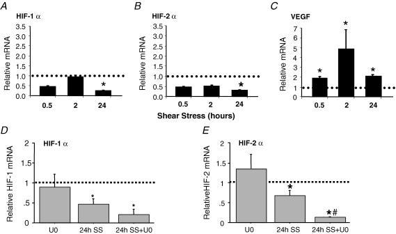
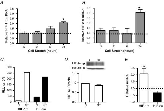
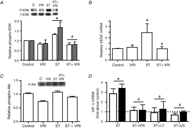
Angiogenesis, which is essential for the physiological adaptation of skeletal muscle to exercise, occurs in response to the mechanical forces of elevated capillary shear stress and cell stretch. Increased production of VEGF is a characteristic of endothelial cells undergoing either stretch- or shear-stress-induced angiogenesis. Because VEGF production is regulated by hypoxia inducible factors (HIFs), we examined whether HIFs play a significant role in the angiogenic process initiated by these mechanical forces. Rat extensor digitorum longus (EDL) muscles were overloaded to induce stretch, or exposed to the dilator prazosin to elevate capillary shear stress, and capillaries from these muscles were isolated by laser capture microdissection for RNA analysis. HIF-1alpha and HIF-2alpha transcript levels increased after 4 and 7 days of stretch, whereas a transient early induction of HIF-1alpha and HIF-2alpha transcripts was detected in capillaries from prazosin-treated muscles. Skeletal muscle microvascular endothelial cells exposed to 10% stretch in vitro showed an elevation in HIF-1alpha and HIF-2alpha mRNA, which was preceded by increases in HIF-binding activity. Conversely, HIF-1alpha and HIF-2alpha mRNA were reduced significantly, and HIF-alpha proteins were undetectable, after 24 h exposure to elevated shear stress (16 dyn cm(-2) (16 x10(-5) N cm(-2)). Given the disparate regulation of HIFs in response to these mechanical stimuli, we tested the requirement of HIF-alpha proteins in stretch- and shear-stress-induced angiogenesis by impeding HIF accumulation through use of the geldanamycin derivative 17-DMAG. Treatment with 17-DMAG significantly impaired stretch-induced, but not shear-stress-induced, angiogenesis. Together, these results illustrate that activation of HIF-1alpha and HIF-2alpha contributes significantly to stretch- but not to shear-stress-induced capillary growth.


Figures

Figure 1
Figure 1. Analysis of mRNA from LCM-captured capillaries shows differential changes in expression of HIF-1α, HIF-2α and VEGF mRNA during overload-induced muscle stretch or prazosin-induced elevation in shear stress
To facilitate the laser capture microdissection (LCM) process, capillaries were visualized by Alexa488-lectin staining (A), then targeted by the laser beam. (B), dark bands on the tissue define individual laser activations. (C), captured capillaries were no longer detectable within the tissue section after removal of the LCM cap. (D), the isolated capillaries were observable on the surface of the LCM cap. (A–D), arrows mark the same three capillaries. Total RNA was isolated from the capillaries captured from extensor digitorum longus (EDL) muscles and the relative mRNA expression levels of HIF-1α (E and H), HIF-2α (F and I), and VEGF-A (G and J), were determined by real-time quantitative PCR (q-PCR). mRNA levels were analysed for samples taken from muscles subjected to stretch for 4 or 7 days (EG), or to elevated shear stress for 2, 4 or 7 days (HJ). In each case, values of mRNA amounts were normalized to 18S rRNA and expressed relative to sham-operated controls. Error bars represent s.e.m.*P < 0.05 versus controls; n = 4.
Figure 2
Figure 2. Effect of 17-DMAG, an Hsp90 inhibitor, on HIF-1α, HIF-2α and VEGF-A protein production in cultured endothelial cells
Cells were treated overnight with cobalt chloride (0.1 mm) to induce HIF protein accumulation, in the absence and presence of 17-DMAG (3 μm). Cell lysates were analysed by Western blot for HIF-1α (A), HIF-2α (B) and VEGF-A (C). Loading variability was normalized according to levels of tubulin (HIF blots) or β-actin (VEGF blots). *P < 0.05 compared with control; #P < 0.05 compared with cobalt chloride treatment; n = 3 or 4 for each protein analysed.
Figure 3
Figure 3. Effect of 17-DMAG on HIF-1α and HIF-2α protein production in rat EDL muscles
A, Western blotting of skeletal muscle extracts showed increased HIF-1α protein in muscles subjected to overload (ST), and this was not observed in muscles subjected to overload in combination with 17-DMAG (ST + DMAG). Ponceau S staining of membranes was used to normalize for loading. *P < 0.05 compared with control unstretched; #P < 0.05 compared with 7 day overload (ST). B, immunostaining was performed on control, 7 day overload (ST), 17-DMAG-treated, and 7 day overload + 17-DMAG-treated (ST+17-DMAG), EDL muscles. Images in the top panels represent HIF-1α immunodetection (green) and those in the bottom panels correspond to HIF-2α immunostaining (green). Corresponding DAPI staining (blue) was used to visualize nuclei. All images were collected using a ×40 objective. Exposure settings used to capture images of HIF-1α staining were identical for all conditions. Pixel intensity quantification of all conditions immunostained for HIF-1α (C) and HIF-2α (D) identified significant increases in staining intensity in overload (ST) compared with control (C; *P < 0.05). Intensity of HIF-α staining in ST+DMAG was reduced significantly compared with ST (#P < 0.05). HIF-1α staining was reduced in unstretched muscles treated with DMAG compared with vehicle treatment ($P < 0.05).
Figure 4
Figure 4. Destabilization of HIF-1α protein using 17-DMAG inhibits stretch-induced but not shear-stress-induced angiogenesis in EDL muscles
Capillary supply was evaluated on the basis of capillary staining by alkaline phosphatase and expressed as capillary-per-fibre ratio, C:F. Rats were treated continuously with vehicle (control) or 17-DMAG delivered locally to the muscle via an implanted osmotic pump. After 14 days, C:F ratio was elevated significantly in stretched muscles compared with control or 17-DMAG-treated muscle (*P < 0.05 versus control). Stretch + 17-DMAG-treated (ST+17-DMAG) muscles had a significantly lower C:F ratio compared with stretch alone (#P < 0.001 versus stretch). 17-DMAG treatment itself did not modify C:F compared with control muscles. Prazosin (PR) treatment for 7 days resulted in significantly greater C:F compared with control. In this case, the C:F for PR+17-DMAG was not different from PR alone. Data are shown as means ± s.e.m.; n = 4.
Figure 5
Figure 5. Regulation of VEGF-A, HIF-1α and HIF-2α by shear-stress stimulation of cultured microvascular endothelial cells
Cells were subjected to 16 dyne cm−2 laminar shear stress (SS) for 0.5, 2 and 24 h. mRNA levels of HIF-1α (A) HIF-2α (B) and VEGF-A (C) were determined by real-time q-PCR. Levels of target genes were normalized to 18S rRNA, then expressed relative to static time-matched controls (set to 1). Each bar represents the mean ± s.e.m. of at least four experiments. *P < 0.05 versus control. In a subsequent set of experiments, endothelial cells were pretreated with vehicle or the MEK1/2 inhibitor U0126 (U0; 0.3 μm) prior to shear or static exposure for 24 h. Real-time q-PCR of HIF-1α mRNA (D) showed a trend for further reductions in HIF-1α mRNA in shear-exposed cells with U0 treatment, while the reduction in HIF-2α mRNA was significantly greater in cells treated with U0 in combination with shear stress than in cells stimulated by shear stress itself (E). For each time point and drug treatment, mRNA levels in sheared samples were expressed as a ratio to their respective static control (set to 1). The dotted line represents the control level for each condition. n = 3; *P < 0.05 compared with control; #P < 0.05 compared with shear stress.
Figure 6
Figure 6. Effect of mechanical stretch on expression of HIF-1α and HIF-2α in cultured microvascular endothelial cells
Cells were subjected to 10% static stretch for 0.5, 2, 6 and 24 h. mRNA levels of HIF-1α (A) and HIF-2α (B) were determined by real-time q-PCR. Levels of target genes were normalized to 18S rRNA and expressed relative to unstretched time-matched controls (set to 1). Each bar represents the mean ± s.e.m. of at least four experiments. *P < 0.05 versus control. Dotted lines represent the level of HIF-1α and HIF-2α mRNA in unstretched samples for each time point. DNA-binding assays were performed on nuclear extracts isolated from control cells and cells subjected to 10% static stretch for 4 h (C). HIF-1α (open bars) and HIF-2α (filled bars) both exhibited greater binding to the hypoxia response element (HRE) in nuclear extracts from stretched cells. Results are representative of three independent experiments. Western blot analysis of HIF-1α from extracts of control or stretched (4 h) endothelial cells showed no significant change in protein quantity in response to stretch (D). Cells were treated with clioquinol (100 μm) or vehicle for 6 h, followed by lysis and analysis of HIF-1α and HIF-2α mRNA transcripts by q-PCR (E). Data are expressed relative to control (set to 1). *P < 0.05 versus control, n = 4.
Figure 7
Figure 7. Effect of VEGFR2-dependent signalling on HIF-1α and HIF-2α gene expression in stretched microvascular endothelial cells
VEGFR2 inhibition (VRI; 10 μm) reduced the typical increase in ERK1/2 phosphorylation (A) induced following 10 min exposure to 10% static stretch (ST). Phospho-ERK values were normalized to total ERK, and expressed relative to unstretched controls. Data are presented as means ± s.e.m.; n = 3 or 4; *P < 0.05 versus control; #P < 0.05 versus ST. VEGFR2 inhibition also reduced the stretch-induced increase in VEGF mRNA observed at 24 h of 10% static stretch (B). Data are presented as means ± s.e.m.; n = 3 or 4; *P < 0.05 versus control; #P < 0.05 versus ST. Phosphorylation of Akt was not altered significantly measured in response to 10 min stretch, in the absence or presence of the VEGFR2 inhibitor (C). Pre-treatment of cells with VEGFR2 inhibitor I (VRI; 10 μm), PI3K inhibitor (LY; 10 μm), or MEK1/2 inhibitor (U0; 0.3 μm), significantly diminished the stretch-induced (ST; 24 h, 10%) expression of HIF-1α (open bars) and HIF-2α mRNA (solid bars) compared with their respective drug-treated unstretched controls (D). Data are presented as means ± s.e.m.; n = 3 or 4; #P < 0.05 versus ST + vehicle (ST).

Similar articles

Cited by

References

    1. Armstrong RB, Ianuzzo CD, Laughlin MH. Blood flow and glycogen use in hypertrophied rat muscles during exercise. J Appl Physiol. 1986;61:683–687. - PubMed
    1. Ausprunk DH, Folkman J. Migration and proliferation of endothelial cells in preformed and newly formed blood vessels during tumor angiogenesis. Microvasc Res. 1977;14:53–65. - PubMed
    1. Berra E, Milanini J, Richard DE, Le Gall M, Vinals F, Gothie E, Roux D, Pages G, Pouyssegur J. Signaling angiogenesis via p42/p44 MAP kinase and hypoxia. Biochem Pharmacol. 2000;60:1171–1178. - PubMed
    1. Bodine SC, Stitt TN, Gonzalez M, Kline WO, Stover GL, Bauerlein R, Zlotchenko E, Scrimgeour A, Lawrence JC, Glass DJ, Yancopoulos GD. Akt/mTOR pathway is a crucial regulator of skeletal muscle hypertrophy and can prevent muscle atrophy in vivo. Nature Cell Biol. 2001;3:1014–1019. - PubMed
    1. Boyd PJ, Doyle J, Gee E, Pallan S, Haas TL. Mitogen-activated protein kinase signaling regulates endothelial cell assembly into networks and the expression of MT1-MMP and MMP—. Am J Physiol Cell Physiol. 2005;288:C659–C668. - PubMed

Publication types

MeSH terms

Substances

LinkOut - more resources