. 2007 Oct;25(10):2677-84.
doi: 10.1634/stemcells.2007-0041. Epub 2007 Jul 12.

Molecular imaging of bone marrow mononuclear cell homing and engraftment in ischemic myocardium

Affiliations

Molecular imaging of bone marrow mononuclear cell homing and engraftment in ischemic myocardium

Ahmad Y Sheikh et al. Stem Cells. 2007 Oct.

Abstract

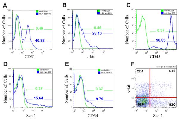
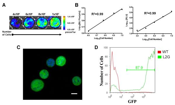
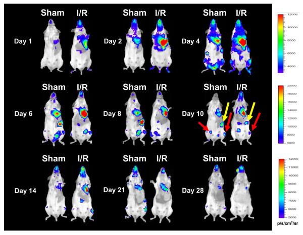
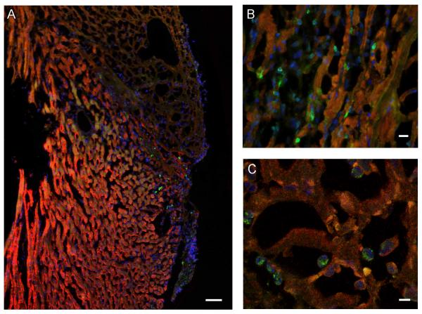
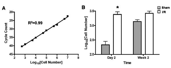
Bone marrow mononuclear cell (BMMC) therapy shows promise as a treatment for ischemic heart disease. However, the ability to monitor long-term cell fate remains limited. We hypothesized that molecular imaging could be used to track stem cell homing and survival after myocardial ischemia-reperfusion (I/R) injury. We first harvested donor BMMCs from adult male L2G85 transgenic mice constitutively expressing both firefly luciferase (Fluc) and enhanced green fluorescence protein reporter gene. Fluorescence-activated cell sorting analysis revealed approximately 0.07% of the population to consist of classic hematopoietic stem cells (lin-, thy-int, c-kit+, Sca-1+). Afterward, adult female FVB recipients (n = 38) were randomized to sham surgery or acute I/R injury. Animals in the sham (n = 16) and I/R (n = 22) groups received 5 x 10(6) of the L2G85-derived BMMCs via tail vein injection. Bioluminescence imaging (BLI) was used to track cell migration and survival in vivo for 4 weeks. BLI showed preferential homing of BMMCs to hearts with I/R injury compared with sham hearts within the first week following cell injection. Ex vivo analysis of explanted hearts by histology confirmed BLI imaging results, and quantitative real-time polymerase chain reaction (for the male Sry gene) further demonstrated a greater number of BMMCs in hearts with I/R injury compared with the sham group. Functional evaluation by echocardiography demonstrated a trend toward improved left ventricular fractional shortening in animals receiving BMMCs. Taken together, these data demonstrate that molecular imaging can be used to successfully track BMMC therapy in murine models of heart disease. Specifically, we have demonstrated that systemically delivered BMMCs preferentially home to and are retained by injured myocardium. Disclosure of potential conflicts of interest is found at the end of this article.


Figures

Figure 1
Figure 1
FACS analysis of bone marrow mononuclear cells (BMMCs) from L2G85 transgenic reporter mice reveals typical proportions of progenitor cells for the FVB strain. Green curves with corresponding percentages representing BMMC samples labeled with (A) CD31, (B) c-kit, (C) CD45, (D) Sca-1, and (E) CD34. Blue curves and percentages demonstrate negative controls (no antibody). (F) Approximately 4.5% of the BMMCs stain positive for both c-kit and Sca-1, similar to preparations used in clinical studies of BMMC transplantation in humans.
Figure 2
Figure 2
Reporter gene activity correlates robustly with cell number. (A) Bioluminescence imaging (BLI) of increasing numbers of BMMC in vitro (total cell count given above corresponding well with color scale bar representing range of signal in p/s/cm2/sr). (B) Correlation of cell numbers (x-axis) with BLI signal (left) and Fluc enzyme activity (right) demonstrate linear relationships with R2 values of 0.99. (C) Confocal laser microscopy of BMMCs demonstrates bright, cytosolic eGFP expression with corresponding nuclei stained blue with DAPI (scale bar = 5 μm). (D) FACS analysis of BMMCs from wild-type control FVB mouse (red) and L2G85 transgenic mouse (green) demonstrates robust eGFP expression by over 87% of the cells.
Figure 3
Figure 3
Bioluminescence imaging demonstrates exogenously delivered BMMC preferentially home to injured myocardium. Images following the same animals (sham on left and I/R injury on right) for 4 weeks following intravenous delivery of L2G85-derived BMMCs (note the maximum values for scale bars in p/s/cm2/sr are different in the three rows). Persistently elevated signal from the area overlying the heart can be observed through day 14, followed by relatively similar decreasing trend in signal intensity by day 28. Images at day 10 demonstrate entrapment of cells in extra-cardiac sites such as the spleen (yellow arrows) and long bones of the lower extremities (red arrows).
Figure 4
Figure 4
Ex vivo imaging confirms presence of transplanted cells within the myocardium. (A) Quantification of signals from ROIs over the thorax demonstrates significantly increased cell numbers in animals with I/R injury (white bars) compared to sham (gray bars) at days 2–6 following transplant (*P<0.05). Mean baseline Log10BLI (p/s/cm2/sr) of unoperated, uninjected mice was 3.3±2.1 (n=5 animals imaged at different time points). This was consistently 1–2 orders of magnitude lower than that of the highest signals attained in the animals receiving cells and I/R injury. (B) Ex vivo imaging of hearts two days following I/R injury with intravenous PBS (left), sham surgery with BMMCs (middle), or I/R injury with BMMCs (right) confirms homing of intravenously delivered BMMCs to the heart (white scale bars = 5 mm).
Figure 5
Figure 5
Histological evaluation confirms BMMC homing to the infarcted heart. Fluorescence microscopy images of a representative heart 2 days following 30 minutes of I/R injury and injection of 5×10⁶ BMMN cells via tail vein. All panels stained with anti-troponin (red), anti-GFP (green), and DAPI (blue). (A) Infarcted area demonstrated by lack of bright troponin stain, with numerous GFP positive cells in the infarct border zone (scale bar = 50 μm). (B) High-power view demonstrating numerous GFP-expressing cells within the myocardium (scale bar = 10 μm). (C) Confocal laser microscopy image confirming presence of GFP-expressing cells within infarcted areas of the myocardium (scale bar = 5 μm).
Figure 6
Figure 6
Real time PCR quantification of surviving male transplanted cells within female hearts. (A) Plot of TaqMan-based Sry-quantification versus number of male cells in female hearts demonstrates a robust correlation between cycle count and known cell number (R2=0.99). (B) Ex vivo TaqMan analysis of hearts undergoing sham surgery (grey bars) versus I/R injury (white bars) demonstrates statistically significant increase of BMMCs homing in to injured hearts compared to sham at day 2 following transplant. This trend persists at week 2, but does not achieve statistical significance (n=5–6/group) (*P<0.05). Both results mirror findings generated by longitudinal bioluminescence imaging in vivo as shown in Figures 3 & 4.
Figure 7
Figure 7
Ventricular function following BMMC therapy. (A) Representative M-mode images of hearts at 4 weeks following I/R injury with administration of PBS (left) versus BMMCs (right) (white scale bars = 5mm). (B) Quantification of left ventricular function demonstrates a trend towards improved functional recovery at 4 weeks post-I/R in animals receiving BMMCs compared to PBS (n=5–6 per time point) but did not achieve statistical significance.

Similar articles

Cited by

References

    1. Orlic D, Kajstura J, Chimenti S, et al. Bone marrow cells regenerate infarcted myocardium. Nature. 2001 Apr 5;410(6829):701–705. - PubMed
    1. Yoon YS, Wecker A, Heyd L, et al. Clonally expanded novel multipotent stem cells from human bone marrow regenerate myocardium after myocardial infarction. J Clin Invest. 2005 Feb;115(2):326–338. - PMC - PubMed
    1. Perin EC, Dohmann HF, Borojevic R, et al. Improved exercise capacity and ischemia 6 and 12 months after transendocardial injection of autologous bone marrow mononuclear cells for ischemic cardiomyopathy. Circulation. 2004 Sep 14;110(11 Suppl 1):II213–218. - PubMed
    1. Strauer BE, Brehm M, Zeus T, et al. Repair of infarcted myocardium by autologous intracoronary mononuclear bone marrow cell transplantation in humans. Circulation. 2002 Oct 8;106(15):1913–1918. - PubMed
    1. Wollert KC, Meyer GP, Lotz J, et al. Intracoronary autologous bone-marrow cell transfer after myocardial infarction: the BOOST randomised controlled clinical trial. Lancet. 2004 Jul 10;364(9429):141–148. - PubMed

Publication types

MeSH terms

Substances