Review
. 2007:422:1-31.
doi: 10.1016/S0076-6879(06)22001-9.

Comparative genomic and protein sequence analyses of a complex system controlling bacterial chemotaxis

Affiliations
Review

Comparative genomic and protein sequence analyses of a complex system controlling bacterial chemotaxis

Kristin Wuichet et al. Methods Enzymol. 2007.

Abstract

Molecular machinery governing bacterial chemotaxis consists of the CheA-CheY two-component system, an array of specialized chemoreceptors, and several auxiliary proteins. It has been studied extensively in Escherichia coli and, to a significantly lesser extent, in several other microbial species. Emerging evidence suggests that homologous signal transduction pathways regulate not only chemotaxis, but several other cellular functions in various bacterial species. The availability of genome sequence data for hundreds of organisms enables productive study of this system using comparative genomics and protein sequence analysis. This chapter describes advances in genomics of the chemotaxis signal transduction system, provides information on relevant bioinformatics tools and resources, and outlines approaches toward developing a computational framework for predicting important biological functions from raw genomic data based on available experimental evidence.


Figures
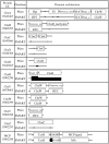
FIG. 1
FIG. 1
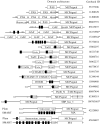
Domain architecture of chemotaxis proteins as visualized in MiST. The MiST database (Ulrich and Zhulin, 2007) uses the domain models from both Pfam and SMART databases. Domains are shown as white boxes with their names inside. Small black, gray, and white boxes indicate predicted transmembrane, low complexity, and signal peptide regions, respectively. The NCBI database GI (GenBank identifier) numbers corresponding to each protein sequence are given under their respective protein names.
FIG. 2
FIG. 2
MCP membrane topology classes. Differing membrane topology divides MCPs into four main classes. (A) Schematic representation of the three-dimensional structure of MCP dimers of different sensor classes. Oval domains are sensory domains of varied secondary structure. Cylinders represent α-helical and coiled coil regions. MCP monomers are differentiated by gray and white coloring. Class I, transmembrane MCPs with extracellular sensory domains; class II, membrane-bound MCPs with N-terminal cytoplasmic sensory domains; class III, membrane-bound MCPs with cytoplasmic sensory domains located C-terminally to the last transmembrane regions (IIIc) or without sensory domains (IIIm); class IV, cytoplasmic MCPs. (B) MCP sensor class can be determined from domain architecture where transmembrane regions and domains are well predicted. Transmembrane regions are indicated by black boxes
FIG. 3
FIG. 3
Diversity of sensory domains in MCPs. All sensory domains are Pfam domain models, except the GAF domain, which is the SMART model (it is slightly longer than the Pfam domain model). HAMP domains are the SMART domain model. MCPs containing hemerythrin and SBP_bac_5 sensory domains represent the atypical topology where the MCP signaling domain is N-terminal of the sensory domain. The Pfam TarH model has shown to be erroneous and will soon be replaced by a correct model termed 4HB_MCP (Ulrich and Zhulin, 2005). Both Pfam and SMART domain architectures are shown for two MCPs with class IIIm membrane topology. Small gray and white boxes indicate predicted low complexity and signal peptide regions, respectively. Black boxes represent transmembrane regions. Long sequences marked by an asterisk (*) were shortened for display and are not to scale.
FIG. 4
FIG. 4
HAMP domain models are imperfect. Both the Pfam SMART HAMP domain models have low sensitivity; however, implementation of both models in MiST enables the identification of HAMP domains in many cases when one of the domain database models misses the target. Note that the Pfam HAMP domain models often (but not always) overlap with one of the transmembrane regions.
FIG. 5
FIG. 5
A common core and diversity of CheA homologs. The domain architectures of selected CheA proteins are shown with their corresponding NCBI GI numbers to the right. All shown domains are from Pfam except for the REC domain (SMART domain model). The dimerization domains shown in gray were delineated by PSI-BLAST analysis; current dimerization domain models have very low sensitivity and fail to predict the domain in many instances. Our analysis shows that the dimerization domain is present in all CheA homologs identified to date (K. Wuichet, unpublished data). Small black, gray, and white boxes indicate predicted transmembrane, low complexity, and signal peptide regions, respectively. The FimL-like domain shows similarity to the FimL pili motility protein, and the Tpt domain shows similarity to Hpt domains, but it has a threonine in place of the conserved histidine (the phosphorylation site). Despite diverse domain architectures, all CheA proteins contain Hpt, dimerization, HATPase_c, and CheW domains, with the latter three forming in a tight protein core. CheA-CheC fusion proteins were also identified; see Fig. 8.
FIG. 6
FIG. 6
The relationship between the domain architecture and the structure of CheA. The domain architecture of the CheA protein directly relates to its structure. The Pfam domain model of CheA (GI 15643465) and its two-dimensional color scheme are shown below the three-dimensional model that has a matching color code. The three-dimensional model consists of three different crystal structures: the Hpt (or P1) domain (PDB identifier 1I5N), the P2 domain (1UOS), and the three core domains (PDB, 1BDJ)—dimerization (or P3) (Pfam, H-kinase_dim), HATPase_c (or P4), and CheW (or P5), respectively, with the linker regions hand drawn. The first two linker regions found in the domain architecture are predicted to be loops between the globular Hpt and P2 domains. The third predicted linker region of CheA suggests that the H-kinase_dim domain model does not capture the entire dimerization domain.
FIG. 7
FIG. 7
Multiple alignment of the P2 domain and its classification. Three subclasses of the P2 domain were identified. A multiple alignment with representative members of each class of P2 domain shows the insertions and deletions that define each class. Positions conserved at 90% or more in an alignment of 116 P2 sequences are shown in gray. Conservation consensus is shown underneath the alignment (h, hydrophobic; l, aliphatic; p, polar; s, small). Black columns show conserved proline and hydrophobic positions in classes I and II. The secondary structure elements are shown above the alignment based on crystal structures from E. coli and T. maritima (McEvoy, 1998; Park, 2004a,b) Black arrows represent β strands. White cylinders represent α helices. Species abbreviations and NCBI GI numbers for each sequence are given at the left (full species name can be found by searching the NCBI nonredundant database with the corresponding GI number).
FIG. 8
FIG. 8
Diversity of CheC homologs. CheC and CheX proteins can be fused to different domains and proteins. Domains shown in gray were missed by the current domain models and were found by PSI-BLAST searches. Their approximate position in corresponding protein sequences is shown. Domain models are from Pfam. Small gray boxes indicate predicted low complexity regions. The NCBI GI number associated with each sequence is shown at the right.
FIG. 9
FIG. 9
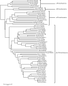
Neighbor-joining tree of the extended CheZ protein family. The CheZ protein family has members present in all classes of Proteobacteria, and the phylogenetic tree suggests its vertical evolution. The sequence identified by a black circle comes from a likely contamination with prokaryotic DNA in the genome of the mosquito Anopheles gambiae.

Similar articles

Cited by

References

    1. Acuna G, Shi W, Trudeau K, Zusman DR. The ‘CheA’ and ‘CheY’ domains of Myxococcus xanthus FrzE function independently in vitro as an autokinase and a phosphate acceptor, respectively. FEBS Lett. 1995;358:31–33. - PubMed
    1. Alexandre G, Zhulin IB. Different evolutionary constraints on chemotaxis proteins CheW and CheY revealed by heterologous expression and protein sequence analysis. J. Bacteriol. 2003;185:544–552. - PMC - PubMed
    1. Alexander RP, Zhulin IB. Submitted for publication. 2007.
    1. Altschul SF, Madden TL, Schaffer AA, Zhang J, Zhang Z, Miller W, Lipman DJ. Gapped BLAST and PSI-BLAST: A new generation of protein database search programs. Nucleic Acids Res. 1997;25:3389–3402. - PMC - PubMed
    1. Anantharaman V, Aravind L. Cache: A signaling domain common to animal Ca(2+)-channel subunits and a class of prokaryotic chemotaxis receptors. Trends Biochem. Sci. 2000;25:535–537. - PubMed

Further Reading

    1. Barnakov AN, Barnakova LA, Hazelbauer GL. Location of the receptor-interaction site on CheB, the methylesterase response regulator of bacterial chemotaxis. J. Biol. Chem. 2001;276:32984–32989. - PubMed
    1. Djordjevic S, Stock AM. Chemotaxis receptor recognition by protein methyl-transferase CheR. Nat. Struct. Biol. 1998;5:446–450. - PubMed
    1. Kall L, Krogh A, Sonnhammer EL. A combined transmembrane topology and signal peptide prediction method. J. Mol. Biol. 2004;338:1027–1036. - PubMed
    1. Lai WC, Hazelbauer GL. Carboxyl-terminal extensions beyond the conserved pentapeptide reduce rates of chemoreceptor adaptational modification. J. Bacteriol. 2005;187:5115–5121. - PMC - PubMed
    1. Letunic I, Copley RR, Pils B, Pinkert S, Schultz J, Bork P. SMART 5.0: Domains in the context of genomes and networks. Nucleic Acids Res. 2006;32:D142–D144. - PMC - PubMed

MeSH terms

LinkOut - more resources