. 2007 Aug;37(8):829-37.
doi: 10.1016/j.ibmb.2007.05.010. Epub 2007 May 29.

Identification and characterization of juvenile hormone esterase gene from the yellow fever mosquito, Aedes aegypti

Affiliations

Identification and characterization of juvenile hormone esterase gene from the yellow fever mosquito, Aedes aegypti

Hua Bai et al. Insect Biochem Mol Biol. 2007 Aug.

Abstract

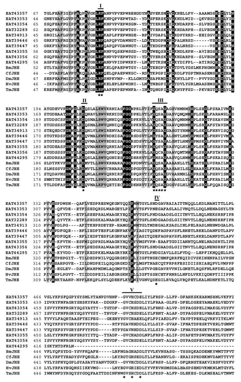
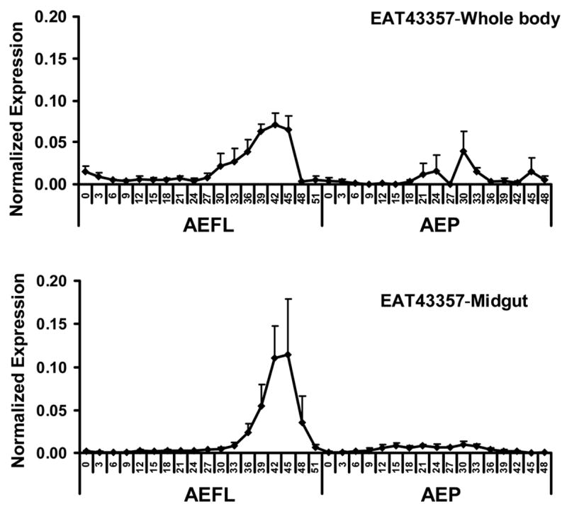
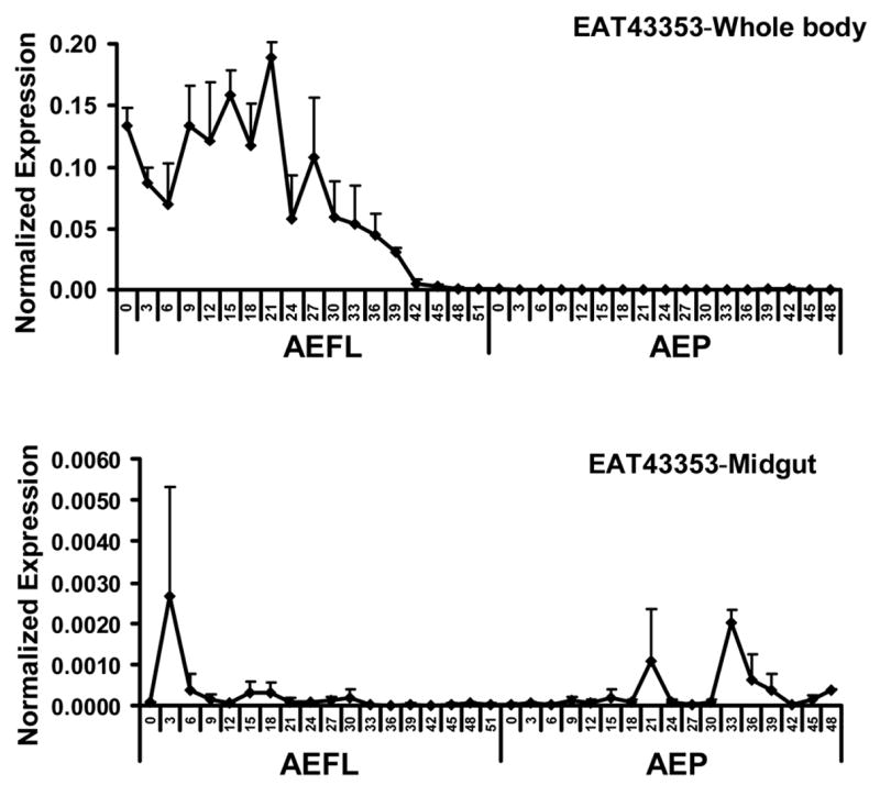
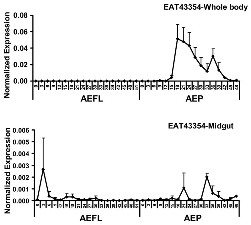
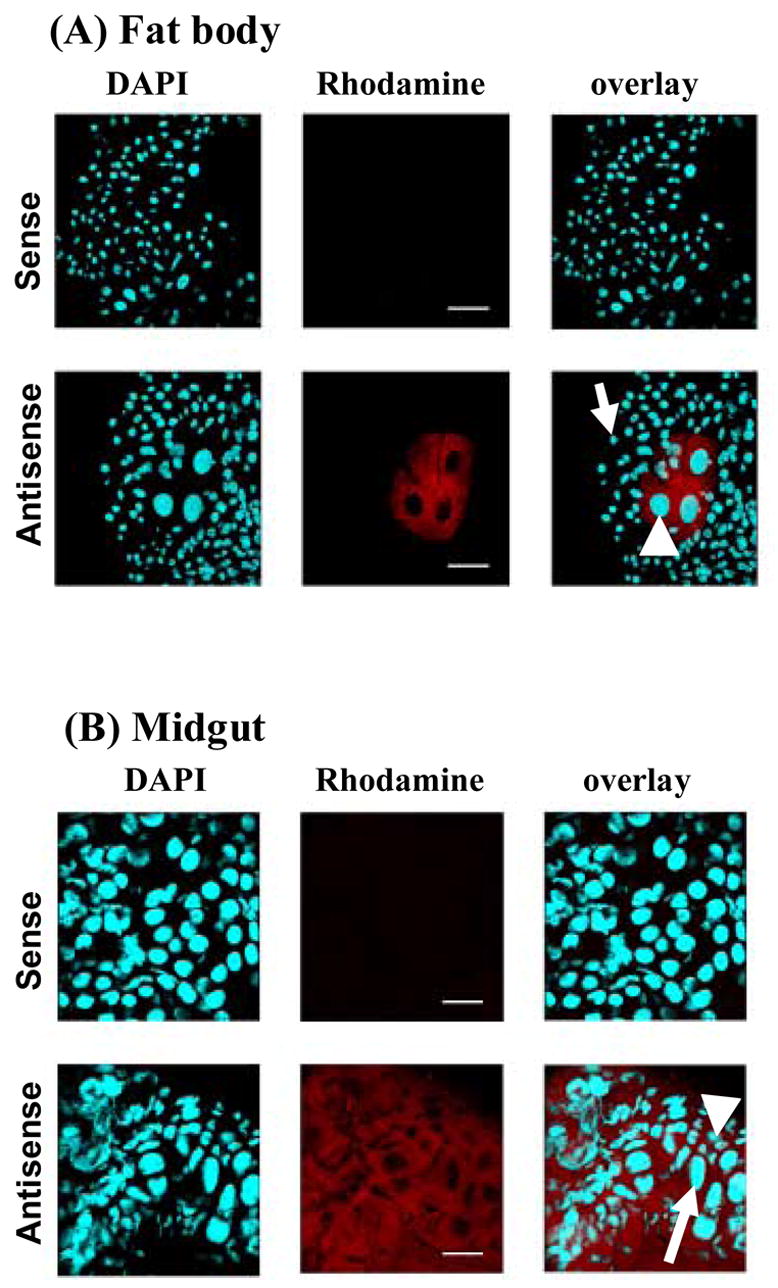
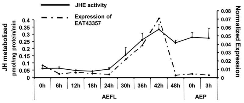
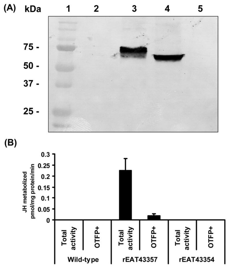
Juvenile hormone esterase (JHE) plays an important role in regulating juvenile hormone titers. Recent sequencing and annotation of the Aedes aegypti genome identified ten putative jhe gene sequences. Analysis of these ten putative jhe gene sequences showed that only three of them, EAT43357, EAT43353 and EAT43354 contained GQSAG motif and showed high sequence similarity with the sequences of jhe genes identified from other insect species. To determine which putative jhe gene(s) code for functional JHE, the mRNA profiles of EAT43357, EAT43353 and EAT43354 were measured during the final instar larval and pupal stages by using quantitative real-time reverse transcriptase polymerase chain reaction (PCR). The mRNA for EAT43357 was detected during the late final instar larval stage. In contrast, EAT43354 mRNA was detected only during the pupal stage and EAT43353 mRNA was detected only during the larval stage. The mRNA of EAT43357 was detected in both fat body and midgut tissues. JHE enzyme levels gradually increased during the final instar larval stage reaching a peak at 42 h after ecdysis into the final instar larval stage. The mRNA expression profiles of EAT43357 correlate with the developmental expression profiles of JHE enzyme activity suggesting that this gene may encode for a functional JHE. The EAT43357 and EAT43354 cDNA were expressed in a baculovirus system. Proteins isolated from Sf9 cells infected with recombinant baculovirus expressing EAT43357 but not EAT43354 gene exhibited JHE activity confirming that EAT43357 gene codes for a functional JHE enzyme.


Figures

Fig. 1
Fig. 1
Alignment of amino acid sequences of ten putative jhe genes from Aedes aegypti, and jhe genes of Drosophila melanogaster (DmJHE, Accession # NM_079034), Choristoneura fumiferana (CfJHE, Accession # AAD34172), Tenebrio molitor (TmJHE, Accession # AAL41023), Heliothis virescens (HvJHE, Accession # AAC38822), Bombyx mori (BmJHE, Accession # AAR37335), using ClustalW program (http://www.ebi.ac.uk/clustalw) and BioEdit Sequence Alignment Editor (http://www.mbio.ncsu.edu/BioEdit/bioedit.html). Five conserved catalytic motifs are indicated by bold lines with the numbers on the top. Numbers on the left show the position of amino acids in each protein. Letters with black and gray shading denote perfect and similar identities, respectively. The motifs important for JHE function are shown as stars. GENSCAN program (http://genome.dkfz-heidelberg.de/cig-bin/GENSCAN/genscan.cgi) was used to predict the full length ORF (547 aa) of EAT43353 gene
Fig. 2
Fig. 2
Developmental profiles of EAT43357 (A), EAT43353 (B) and EAT43354 (C) mRNA in whole body homogenates and midgut. Newly molted 4th instar larvae were sampled every 3 hr until pupation. Newly molted pupae were sampled every 3 hr until adult emergence. The pharate pupae with tanned respiratory trumpets appearing at 48 hr and 51hr AEFL were also staged. The mRNA levels of these genes were normalized using S7RP ribosomal RNA as an internal standard. Mean ± SE for three independently staged sets of larvae and pupae are shown. AEFL, after ecdysis into the final instar larval stage; AEP, after ecdysis into the pupa stage.
Fig. 2
Fig. 2
Developmental profiles of EAT43357 (A), EAT43353 (B) and EAT43354 (C) mRNA in whole body homogenates and midgut. Newly molted 4th instar larvae were sampled every 3 hr until pupation. Newly molted pupae were sampled every 3 hr until adult emergence. The pharate pupae with tanned respiratory trumpets appearing at 48 hr and 51hr AEFL were also staged. The mRNA levels of these genes were normalized using S7RP ribosomal RNA as an internal standard. Mean ± SE for three independently staged sets of larvae and pupae are shown. AEFL, after ecdysis into the final instar larval stage; AEP, after ecdysis into the pupa stage.
Fig. 3
Fig. 3
Tissue specific expression of EAT43357 at 45 hr AEFL in the fat body (A) and midgut (B). In situ hybridization was performed on whole mounts of fat body and midgut tissues using a DIG labeled antisense mRNA probe for EAT43357. Nuclear staining was performed with DAPI. The transcripts were localized using anti-DIG Rhodamine conjugate. Sense strand RNA probes were used as the negative control shown in the top panel of each tissue. No gene expression was detected in the negative control panels. (A). EAT43357 mRNA was detected in fat body cells (white arrowhead), but not in the epidermal cells (white arrow); (B). EAT43357 mRNA was detected in both large larval (white arrow) and small imaginal midgut cells (white arrowhead). Scale bar: 100 μm.
Fig. 4
Fig. 4
Developmental profile of JHE enzymatic activity. JHE activity (solid line) was determined in whole body extracts collected at 6 hr intervals during the final instar larval stage and early pupal stage. For each time point, five larvae or pupae were collected. Mean ± SE for three independently staged sets of larvae and pupae are shown. The mRNA expression profile of EAT43357 is shown as a dashed line.
Fig. 5
Fig. 5
(A) Western blot analysis of the product of EAT43357 cDNA expressed in Sf9 cells. Lane 1: Protein molecular weight marker; Lane 2: Crude protein extract from cells infected with wild-type AcMNPV; Lane 3: Crude protein extract from cells infected with AcMNPV expressing rEAT43357;. Lane 4: Crude protein extract from cells infected with AcMNPV expressing rEAT43354; Lane 5: Crude protein extract from cells infected with AcMNPV expressing rEAT43353. (B) The JHE activity of rEAT43357 protein expressed using the baculovirus system. The total activity bar represents the JHE activity of total proteins, while OTFP+ bar represents the JHE activity when total proteins were incubated with the JHE inhibitor, OTFP. Cell lysates from wild-type baculovirus infected Sf9 cell were used as a negative control. Mean ± SE for three independently expressed recombinant proteins are shown.

Similar articles

Cited by

References

    1. Bonning BC, Hoover K, Booth TF, Duffey S, Hammock BD. Development of a recombinant baculovirus expressing a modified juvenile hormone esterase with potential for insect control. Arch Insect Biochem Physiol. 1995;30:177–194.
    1. Bradford MM. Rapid and sensitive method for quantitation of microgram quantities of protein utilizing principle of protein dye binding. Anal Biochem. 1976;72:248–254. - PubMed
    1. Browder MH, D’Amico LJ, Nijhout HF. The role of low levels of juvenile hormone esterase in the metamorphosis of Manduca sexta. J Insect Sci. 2001;1:11. - PMC - PubMed
    1. Campbell PM, Harcourt RL, Crone EJ, Claudianos C, Hammock BD, Russell RJ, Oakeshott JG. Identification of a juvenile hormone esterase gene by matching its peptide mass fingerprint with a sequence from the Drosophila genome project. Insect Biochem Mol Biol. 2001;31:513–520. - PubMed
    1. Campbell PM, Healy MJ, Oakeshott JG. Characterization of juvenile hormone esterase in Drosophila melanogaster. Insect Biochem Mol Biol. 1992;22:665–677. - PubMed

Publication types

MeSH terms

LinkOut - more resources