. 2007 Aug 15;308(2):534-46.
doi: 10.1016/j.ydbio.2007.06.011. Epub 2007 Jun 21.

Delta and Egfr expression are regulated by Importin-7/Moleskin in Drosophila wing development

Affiliations

Delta and Egfr expression are regulated by Importin-7/Moleskin in Drosophila wing development

Alysia D Vrailas-Mortimer et al. Dev Biol. .

Abstract

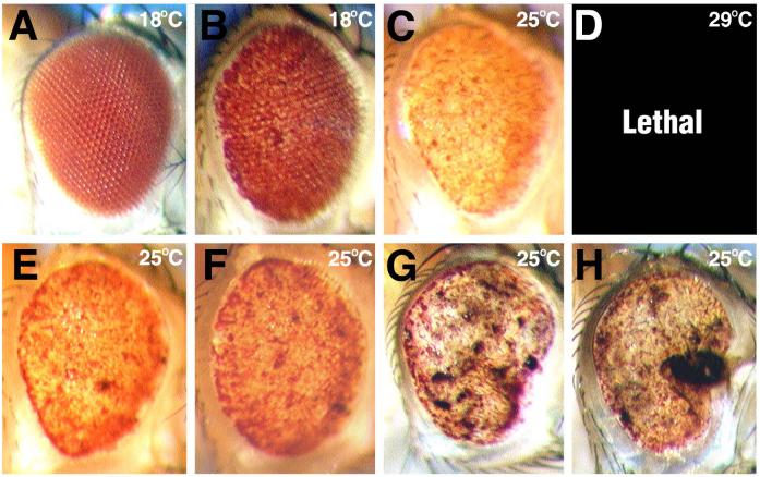
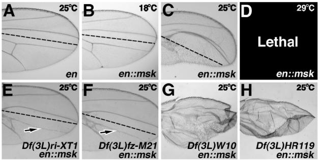
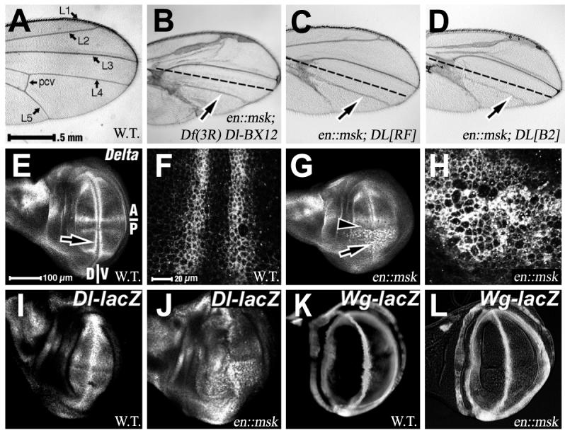
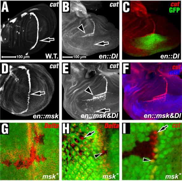
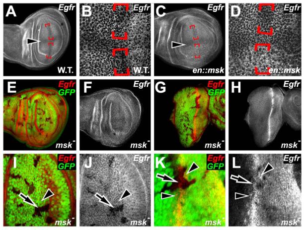
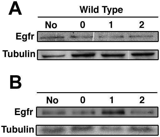
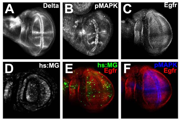
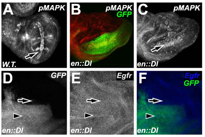
Drosophila DIM-7 (encoded by the moleskin gene, msk) is the orthologue of vertebrate Importin-7. Both Importin-7 and Msk/DIM-7 function as nuclear import cofactors, and have been implicated in the control of multiple signal transduction pathways, including the direct nuclear import of the activated (phosphorylated) form of MAP kinase. We performed two genetic deficiency screens to identify deficiencies that similarly modified Msk overexpression phenotypes in both eyes and wings. We identified 11 total deficiencies, one of which removes the Delta locus. In this report, we show that Delta loss-of-function alleles dominantly suppress Msk gain-of-function phenotypes in the developing wing. We find that Msk overexpression increases both Delta protein expression and Delta transcription, though Msk expression alone is not sufficient to activate Delta protein function. We also find that Msk overexpression increases Egfr protein levels, and that msk gene function is required for proper Egfr expression in both developing wings and eyes. These results indicate a novel function for Msk in Egfr expression. We discuss the implications of these data with respect to the integration of Egfr and Delta/Notch signaling, specifically through the control of MAP kinase subcellular localization.


Figures

Figure 1
Figure 1. Msk over-expression affects eye development
(A-H) show adult eyes, anterior right, dorsal up, genotypes indicated bottom right. Temperatures raised are indicated top right. (A) GMR:GAL4 alone shows phenotypically wild type eye at lower temperatures. (B-D) Eye phenotypes of Msk over-expression posterior to the morphogenetic furrow (GMR::msk) at increasing temperatures. (E-F) Suppression of GMR::msk eye phenotype at 25°C by loss of indicated genomic deficiencies. (G-H) Enhancement of GMR::msk eye phenotype at 25°C by loss of indicated genomic deficiencies.
Figure 2
Figure 2. Msk over-expression affects wing development
(A-H) show adult wings, anterior up, distal right, genotypes indicated bottom right. Dotted lines in each panel represent the anterior/posterior (A/P) boundary. Temperatures raised are indicated top right. (A) en:GAL4 alone shows phenotypically wild type wing at lower temperatures. (B-D) Adult wing phenotypes of Msk over-expression within posterior compartments of developing wings (en::msk) at increasing temperatures. (E-F) Suppression of en::msk wing phenotype at 25°C by loss of indicated genomic deficiencies. Arrows indicate rescue of L4 vein. (G-H) Enhancement of en::msk wing phenotype at 25°C by loss of indicated genomic deficiencies.
Figure 3
Figure 3. Loss-of-function Delta mutations suppress Msk over-expression phenotypes in adult wings
(A-D) Adult wings, anterior up, distal right. Magnification equal in (A-D) and (E, G, I-L) and (F and H) as indicated. Genotypes indicated below right. (A) The normal venation pattern is labeled with longitudinal veins (L1-L5) and posterior crossvein (pcv) indicated. (B) Dotted line separates anterior (top) from posterior (bottom) of the wing in this and subsequent panels. Msk over-expression is driven in the posterior compartment (en::msk) of genomic deficiency Df(3R) Dl-BX12. Arrow indicates rescue of L4 vein normally absent in en::msk wings. (C-D) Arrows indicates suppression of en::msk by loss-of-function in one copy of DeltaRF (C), and DeltaB2 (D). (E-L) Larval wings, anterior/posterior (A/P) and dorsal/ventral (D/V) as indicated in (E). Antigens shown indicated above right. Genotypes indicated below right. (E-F) Wild type Delta protein expression. (F) shows high magnification of the area at arrow in (E). (G-H) Msk over-expression in the posterior compartment (en::msk). (G) Arrowhead shows A/P boundary. Arrow indicates increased Delta protein expression. (H) shows high magnification of the area at arrow in (G). (I-L) anti-β-Galactosidase expression in (I) Delta-lacZ, (J) Delta-lacZ, en::msk, (K) Wingless-lacZ, and (L) Wingless-lacZ, en::msk wing discs.
Figure 4
Figure 4. Msk over-expression alters Dl protein expression
(A-G, and I) Larval wings, anterior up, dorsal right. (H) Larval eye, anterior right. Antigens shown and GFP indicated above right. Genotypes indicated below right. (A) Wild type Cut protein expression in larval wing. Arrow indicates posterior compartment Cut expression. (B) Delta over-expression in the posterior compartment (en::Dl). Arrow indicates loss of Cut protein expression in the posterior compartment (compare to arrow in (A)). Arrowhead indicates ectopic Cut protein expression along anterior/dorsal cells. (C) Shows Delta expression (marked by posterior GFP expression) merged with ectopic Ct protein expression in adjacent anterior/dorsal cells. (D) Msk over-expression in the posterior compartment (en::msk). Arrow indicates reduced Cut protein expression in the posterior compartment. (E) Over-expression of both Msk and Delta in posterior wing disc compartments (en:GAL4 driving both UAS:Delta and UAS:msk, en::Dl, en::msk). Arrow shows ectopic cut expression in posterior/dorsal cells. (F) Shows Msk protein expression (in blue) with Cut protein expression (in red) in both anterior/dorsal cells adjacent to Msk/Dl co-expressing cells, as well as in posterior/dorsal cells not adjacent to Msk/Dl co-expressing cells. (G-I) Mosaic msk5 null somatic clones in late third instar larval wing discs (G, I) and eye discs (H). msk5 null clones are negatively marked with GFP (green) in all panels. (G) Delta protein expression in msk5 null clones is not significantly altered within or outside clones in developing wings. (H) Delta protein expression in eyes with msk5 null clones is not significantly altered within or outside clones either within the morphogenetic furrow (arrowhead) or posterior to the morphogenetic furrow (arrow). (I) Cut protein expression in wings with msk5 null clones is not significantly altered within (arrow) or outside (arrowhead) the clones. Magnification is equal in (A and D), (B, C, E, F) and (G, H) as indicated.
Figure 5
Figure 5. Egfr expression is altered in msk gain- and loss-of-function
Panels show late third instar larval wings (A-D, E-F, I-J) anterior up, dorsal left, and eyes (G-H, and K-L) anterior right. (A-B) Normal Egfr protein expression in wing discs. (A) Arrowhead denotes the Anterior/Posterior compartment boundary. (B) shows high magnification of the area at arrowhead in (A). (C-D) shows Egfr expression in en::msk wing discs. (C) Arrowhead denotes the Anterior/Posterior boundary. Msk expression occurs in the posterior compartment. Note the increased Egfr protein expression within this compartment. (D) shows high magnification of the area at arrowhead in (C). (E-L) All panels show msk5 null clones marked by the absence of GFP expression (green) within the tissue. All panels show Egfr protein expression (red or white as appropriate) within tissues. Note the decreased Egfr expression within clones in the developing wing (E-F) and eyes (G-H). (I-J) shows high magnification of clones in developing wings. Note Egfr protein expression is decreased within clones (arrow) as compared to outside clones (arrowhead). (K-L) shows high magnification of clones in developing eyes. Note Egfr protein expression is decreased within clones (arrow) as compared to outside clones (arrowhead).
Figure 6
Figure 6. Up-regulation of Egfr by Msk is transient
Western Blot of wild type and hs:msk whole larval extracts probed with anti-Egfr and anti-tubulin antibodies. (A) Egfr expression in wild type larvae after no heat shock (No), a one hour heat shock followed by no recovery (0), 1 hour recovery (1) and 2 hours of recovery (2). (B) Egfr expression in hs:msk larvae after no heat shock (No), a one hour heat shock followed by no recovery (0), 1 hour recovery (1) and 2 hours of recovery (2). Note increased Egfr expression after 1 hour of recovery. Tubulin is protein loading control.
Figure 7
Figure 7. Delta and Egfr expression are specific to MAPK subcellular localization
All panels show wild type late third instar larval wings, anterior up, dorsal left. (A) Delta expression in developing wings. (B) pMAPK expression in developing wings. (C) Egfr expression in developing wings. (D) MAPK/GAL4 (MG)-driven GFP expression to show nuclear MAPK in developing wings. (E) Egfr (red) co-localized with MAPK/GAL4 (MG)-driven GFP (green). (F) Egfr (red) co-localized with pMAPK expression (blue).
Figure 8
Figure 8. Delta expression induces pMAPK expression without increasing Egfr expression
All panels show wild type late third instar larval wings, anterior up, dorsal left. (A) pMAPK expression in a normal wing disc. Arrow indicates expression in veins and wing margin in the posterior compartment. (B-F) Delta over-expression in the posterior compartment (en::Dl). GFP shows where ectopic Dl is expressed in (B). Arrow in (C) denotes increased pMAPK expression in the posterior compartment of these discs. (D-F) Egfr expression in en::Dl discs. Arrows denote anterior expression in all panels. Arrowheads denote posterior expression in all panels. Dl expression is marked by GFP in (D) and (F).

Similar articles

Cited by

References

    1. Artavanis-Tsakonas S, Matsuno K, Fortini ME. Notch signaling. Science. 1995;268:225–232. - PubMed
    1. Artavanis-Tsakonas S, Rand MD, Lake RJ. Notch signaling: cell fate control and signal integration in development. Science. 1999;284:770–776. - PubMed
    1. Baker NE. Transcription of the segment-polarity gene wingless in the imaginal discs of Drosophila, and the phenotype of a pupal-lethal wg mutation. Development. 1988;102:489–497. - PubMed
    1. Baker SE, Lorenzen JA, Miller SW, Bunch TA, Jannuzi AL, Ginsberg MH, Perkins LA, Brower DL. Genetic interaction between Integrins and moleskin, a gene encoding a Drosophila homolog of Importin-7. Genetics. 2002;162:285–296. - PMC - PubMed
    1. Baron M. An overview of the Notch signalling pathway. Semin Cell Dev Biol. 2003;14:113–9. - PubMed

Publication types

MeSH terms

LinkOut - more resources