. 2007 Aug 15;308(2):507-19.
doi: 10.1016/j.ydbio.2007.06.007. Epub 2007 Jun 13.

Mago Nashi and Tsunagi/Y14, respectively, regulate Drosophila germline stem cell differentiation and oocyte specification

Affiliations

Mago Nashi and Tsunagi/Y14, respectively, regulate Drosophila germline stem cell differentiation and oocyte specification

David H Parma et al. Dev Biol. .

Abstract

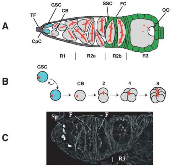
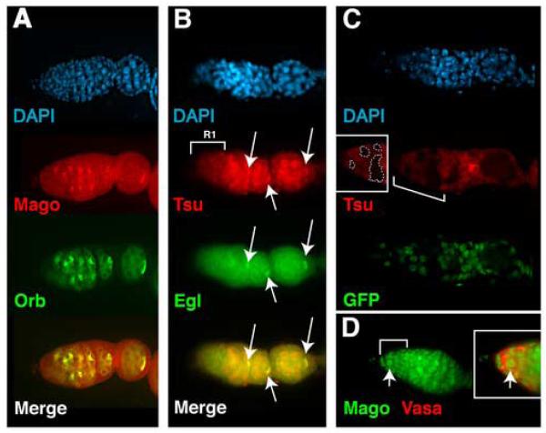
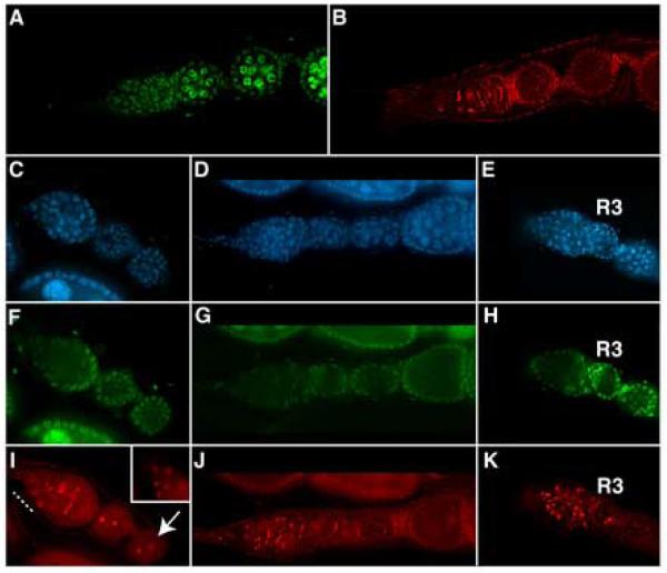
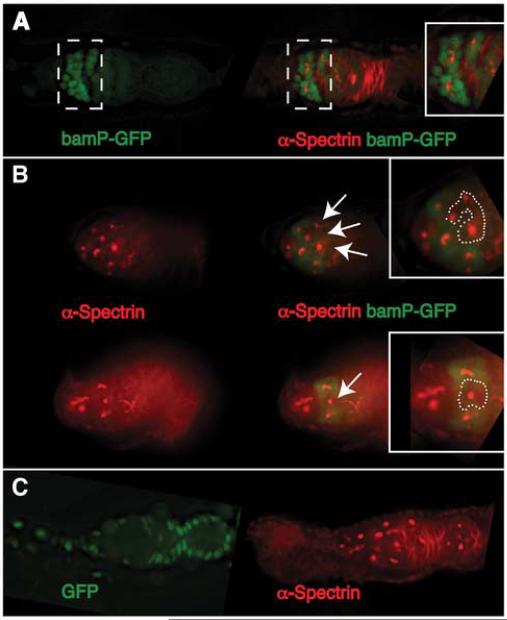
A protein complex consisting of Mago Nashi and Tsunagi/Y14 is required to establish the major body axes and for the localization of primordial germ cell determinants during Drosophila melanogaster oogenesis. The Mago Nashi:Tsunagi/Y14 heterodimer also serves as the core of the exon junction complex (EJC), a multiprotein complex assembled on spliced mRNAs. In previous studies, reduced function alleles of mago nashi and tsunagi/Y14 were used to characterize the roles of the genes in oogenesis. Here, we investigated mago nashi and tsunagi/Y14 using null alleles and clonal analysis. Germline clones lacking mago nashi function divide but fail to differentiate. The mago nashi null germline stem cells produce clones over a period of at least 11 days, suggesting that mago nashi is not necessary for stem cell self-renewal. However, germline stem cells lacking tsunagi/Y14 function are indistinguishable from wild type. Additionally, in tsunagi/Y14 null germline cysts, centrosomes and oocyte-specific components fail to concentrate within a single cell and oocyte fate is not restricted to a single cell. Together, our results suggest not only that mago nashi is required for germline stem cell differentiation but that surprisingly mago nashi functions independently of tsunagi/Y14 in this process. On the other hand, Tsunagi/Y14 is essential for restricting oocyte fate to a single cell and may function with mago nashi in this process.


Figures

Fig. 1
Fig. 1
Development of a wild-typeDrosophila germarium. In all figures, anterior is to the left and posterior to the right. (A) Germline stem cells (GSC; blue) are found at the anterior end of the germarium (region 1; R1), within the stem cell niche. In addition to the GSC, the stem cell niche contains the cap cells (CpC) that anchor the GSC to the niche. The anterior-most cells are somatic terminal filament (TF) cells. As germline cells (gray) develop, they move posteriorly within R1, becoming cystoblasts (CB) that undergo four incomplete divisions to form a 16-cell cyst. Region 2 (consisting of R2a and R2b) contains 16-cell cysts that are no longer mitotically active. Spectrosomes (red) are found in GSCs and CBs, and develop into fusomes (red). In R2b, somatic stem cells (SSC; green) produce follicle cells (FC; green) that encapsulate 16-cell cysts. Region 3 (R3) contains the stage 1 egg chamber, consisting of follicle cells, 15 germline nurse cells and an oocyte at the posterior end (OO; white). (B) The transition of a GSC to a CB and subsequent formation of a 2, 4 and 8-cell cyst. Both spectrosomes and fusomes are depicted in red. (C) A confocal micrograph illustrating development of the fusome; spectrosome (Sp) and fusome (F).
Fig. 2
Fig. 2
Mago and Tsu/Y14 are detected within nuclei in the germarium and, in the cytoplasm, colocalize with Orb and Egl. (A) Cytoplasmic Mago colocalizes with Orb throughout the germarium. (B) Tsu accumulates within nuclei and colocalizes with Egl within the cytoplasm throughout the germarium. The mouse monoclonal α-Tsu, Tsu208, was employed to determine the distribution of Tsu/Y14. The arrows point to cytoplasmic colocalization of Tsu/Y14 and Egl in the germarium. (C) Germline clones of tsuΔ3-5, illustrating the expression of Tsu/Y14 in R1. Clones are identified both by the absence of detectable Tsu and the absence of detectable Hist2avD-GFP. The bracketed area is enhanced and shown within the insert (tsuΔ3-5 clones are outlined by the white dotted lines). (D) Detection of GFP-Mago in nuclei throughout R1 and R2. Detection of cytoplasmic Vasa (red) confirms the germline accumulation of GFP-Mago within the nucleus of a GSC (indicated by the arrows). The bracketed area is magnified and illustrated in the insert.
Fig. 3
Fig. 3
Homozygous magoSHL-1 germline cells are blocked in early germarial development but homozygous tsuΔ3-5 cells block at a post-germarial stage. In all panels, anti-α-Spectrin antibody (α-3A9, red) marks spectrosomes and fusomes, DAPI staining (blue) identifies DNA and Hist2avD-GFP (green) marks non-mosaic cells. (A,B) The expression of the Hist2avD-GFP transgene and α-Spectrin staining, respectively, are illustrated in the germarium and stage 1 egg chamber of a non-mosaic ovariole. (C,F,I) An ovariole containing homozygous magoSHL-1 germline cells (GFP) is depicted. In (I) it can be seen that homozygous magoSHL-1 cells contain spectrosome-like organelles in germarial R1 (marked by dashed white line and magnified within insert), R2, R3 and in pseudo-egg chambers (arrow). (D,G,J) Homozygous tsuΔ3-5 germline cells develop to stage 3-4. Fusome development in homozygous tsuΔ3-5 appears normal. (E,H,K) Double mutant germline clones homozygous for tsuΔ3-5, magoSHL-1 contain spectrosome-like organelles in germarial regions 1, 2 and 3.
Fig. 4
Fig. 4
Germline stem cell differentiation requires mago+. In all panels anti-α-Spectrin (α-3A9) staining identifies spectrosomes and fusomes (red). (A) On the left, a non-mosaic germarium carrying bamP-GFP, shows the transcription of bamP-GFP (green). On the right, the same germarium is shown, illustrating the normal development of fusomes from spectrosomes (red) and transcription of bamP-GFP. The bracketed region is magnified and shown in the box at the right. (B) The pattern of α-Spectrin staining (left) and transcription of bam-GFP (right) in germaria containing magoSHL-1 germline clones is illustrated. Cells displaced from the stem cell niche that contain spectrosome-like organelles (homozygous magoSHL-1 germline clones) and in which transcription of bam-GFP is silenced are identified by arrows (magnified within the insert and outlined by white dotted lines). (C) Double mutant magoSHL-1 bgcn1 germline clones are indistinguishable from magoSHL-1. On the left, the absence of Hist2avD-GFP transgene expression is used to identify magoSHL-1 bgcn1 clones.
Fig. 5
Fig. 5
Somatic stem cell division and differentiation to produce follicle cells does not require mago+ function. Homozygous magoSHL-1 somatic stem cells and their progeny are identified by the absence of Hist2avD-GFP. To identify ovarian somatic cells, antibodies immunoreactive with the Traffic jam protein (α-TJ; the Drosophila melanogaster homolog of the retroviral oncoprotein v-Maf8 and vertebrate large MAF transcription factors) were used (Li, et al., 2003). Ovariole development is indistinguishable from wild type up to at least S9. Part of the bracketed region is within the insert, containing an enhanced and magnified image to illustrate that the oocyte nucleus has migrated to the anterior pole of the oocyte. The dotted lines outline the ovariole, showing the position of the somatic cells. Red arrows, in the merged image, point to the position where the dotted line starts in the GFP image above it.
Fig. 6
Fig. 6
Accumulation of Orb protein in homozygous tsuΔ3-5 germline clones is abnormal. In the panels, nuclei are marked with DAPI (blue), non-mosaic cells express the Hist2avD-GFP transgene (green) and α-Orb (red) allows monitoring of the accumulation of Orb protein. (A,B,C) Accumulation of Orb in a non-mosaic ovariole is shown. (C) Orb is first detected in R2A (bracketed and magnified within insert). Orb can also be detected within the posterior pole of a stage 1 (S1) and 5 (S5) egg chamber. (D,E,F) Normal accumulation of Orb is not detectable in homozygous tsuΔ3-5 germline clones. (F) In homozygous tsuΔ3-5 both the accumulation and distribution of Orb are abnormal when compared to non-mosaic germline cells (arrows indicate non-mosaic egg chambers).
Fig. 7
Fig. 7
Restriction of oocyte fate to a single cell requires tsu+ function within the germline. Staining of DNA is visualized by DAPI (blue), staining of a component of the synaptonemal complex (SC) is observed employing α-C(3)G antibodies (red), and the Hist2avD-GFP transgene is used to distinguish non-mosaic cells (green) from homozygous tsuΔ3-5 cells (GFP). In all panels, the germarium (g) is to the left of the dotted, white, vertical line. (B,E) The arrows point to cysts in which SC formation has occurred within multiple nuclei of a cyst and the arrowheads point to cysts in which a single nucleus, the oocyte nucleus, containing SC. (A,B) A wild-type ovariole illustrating SC formation. In region 2a, cysts of wild-type germaria, first the two pro-oocytes enter meiosis and form SC. Subsequently the two cells with 3 ring canals also enter meiosis and form SC. In region 2b, only the two pro-oocytes retain SC and within R3, SC is detected only in the oocyte. (C,D,E) An ovariole with homozygous tsuΔ3-5 germline clones in the germarium and heterozygous egg chambers outside of the germarium. Unlike wild-type germline cells, four cells retain SC in region 2b and 3. Retention of SC in homozygous tsuΔ3-5 germline cells throughout germarial development suggests that oocyte differentiation is not restricted to a single cell.
Fig. 8
Fig. 8
Centrosomes fail to accumulate within a single cell in tsuΔ3-5 germline germarial cells. The centromsomal protein, CP309, is visualized employing α-CP309. (A) In wild-type germaria (g), centrosomes accumulate within a single cell, the oocyte (indicated by the arrows). (B) In tsuΔ3-5 clonal germaria, the centrosomes do not concentrate in a single cell.

References

    1. Andersen CB, et al. Structure of the exon junction core complex with a trapped DEAD-box ATPase bound to RNA. Science. 2006;313:1968–1972. - PubMed
    1. Ballut L, et al. The exon junction core complex is locked onto RNA by inhibition of eIF4AIII ATPase activity. Nat. Struct. Mol. Biol. 2005;12:861–869. - PubMed
    1. Bolivar J, et al. Centrosome migration into the Drosophila oocyte is independent of BicD and egl, and of the organisation of the microtubule cytoskeleton. Development. 2001;128:889–1897. - PubMed
    1. Bono F, et al. The crystal structure of the exon junction complex reveals how it maintains a stable grip on mRNA. Cell. 2006;126:713–725. - PubMed
    1. Boswell RE, et al. Mutations in a newly identified Drosophila melanogaster gene, mago nashi, disrupt germ cell formation and result in the formation of mirror-image symmetrical double abdomen embryos. Development. 1991;113:373–384. - PubMed

Publication types

MeSH terms

LinkOut - more resources