. 2007 Sep;207(1):64-74.
doi: 10.1016/j.expneurol.2007.05.028. Epub 2007 Jun 14.

Reinnervation of the tibialis anterior following sciatic nerve crush injury: a confocal microscopic study in transgenic mice

Affiliations

Reinnervation of the tibialis anterior following sciatic nerve crush injury: a confocal microscopic study in transgenic mice

Christina K Magill et al. Exp Neurol. 2007 Sep.

Abstract

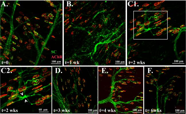
Transgenic mice whose axons and Schwann cells express fluorescent chromophores enable new imaging techniques and augment concepts in developmental neurobiology. The utility of these tools in the study of traumatic nerve injury depends on employing nerve models that are amenable to microsurgical manipulation and gauging functional recovery. Motor recovery from sciatic nerve crush injury is studied here by evaluating motor endplates of the tibialis anterior muscle, which is innervated by the deep peroneal branch of the sciatic nerve. Following sciatic nerve crush, the deep surface of the tibialis anterior muscle is examined using whole mount confocal microscopy, and reinnervation is characterized by imaging fluorescent axons or Schwann cells (SCs). One week following sciatic crush injury, 100% of motor endplates are denervated with partial reinnervation at 2 weeks, hyperinnervation at 3 and 4 weeks, and restoration of a 1:1 axon to motor endplate relationship 6 weeks after injury. Walking track analysis reveals progressive recovery of sciatic nerve function by 6 weeks. SCs reveal reduced S100 expression within 2 weeks of denervation, correlating with regression to a more immature phenotype. Reinnervation of SCs restores S100 expression and a fully differentiated phenotype. Following denervation, there is altered morphology of circumscribed terminal Schwann cells demonstrating extensive process formation between adjacent motor endplates. The thin, uniformly innervated tibialis anterior muscle is well suited for studying motor reinnervation following sciatic nerve injury. Confocal microscopy may be performed coincident with other techniques of assessing nerve regeneration and functional recovery.


Figures

Figure 1
Figure 1
A. Confocal imaging of the tibialis anterior shows that motor endplates are singly innervated by terminal axons in uninjured adult mice. Axons are constitutively labeled with YFP in thy1-YFP(16) mice, while Alexa 594-bungarotoxin labeled acetylcholine receptors delineate motor endplates. B One week after sciatic nerve crush, all motor endplates are denervated, but YFP persists during the process of Wallerian degeneration. Therefore, some terminal axons contact motor endplates, but proximally there is a lack of axonal continuity. C Two weeks after injury, less YFP-labeled axonal debris remains. Although the majority of motor endplates remain denervated, a few rapidly regenerating terminal axonal sprouts are noted. D Three weeks after injury, all endplates are singly or dually innervated, but there are also a number of unmatched, disorganized terminal axons. E Four weeks after injury, the majority of endplates are singly innervated with very few unmatched terminal axons. F Six weeks after injury, all terminal axons are matched and all motor endplates are singly innervated.
Figure 2
Figure 2
A. The percentage of endplates that are either denervated, or reinnervated with one or two axons is shown before crush injury and up to 6 weeks after injury. B. Functional recovery following sciatic crush injury is correlated to the sciatic functional index (SFI) and print length factor (PLF). Weekly statistically significant improvements (p<0.05) are noted starting three weeks after crush injury and approximate pre-injury levels by 6 weeks following crush injury.
Figure 3
Figure 3
A. Confocal image of uninjured tibialis anterior muscle demonstrates mature peripheral SCs and TSCs. B. One week after crush injury, there are qualitatively fewer TSCs expressing S100, and those that are present are less bright. Peripheral SCs possess less uniform morphology, and express GFP above baseline levels. C1. Two weeks later, TSCs are less bright. TSCs appear to extend processes that interconnect multiple motor endplates in a pattern not observed in the uninjured state or after complete recovery. C2. Magnification of the inset outlined in C1. demonstrates the proliferation of peripheral SCs proximal to the motor endplates at two weeks following crush injury (see arrows). D. Three weeks later, GFP expression by TSCs is drastically increased. E. Four weeks later, both peripheral and TSCs remain brightly labeled. F. Six weeks later, SC distribution, morphology, and GFP expression approximates the pre-injury state.
Figure 4
Figure 4
A. Thy1-CFP(23)/S100-GFP mice combine the SC and terminal axon characteristics seen in single transgenic mice. Quantitative assessment, however, is exclusively performed on the single transgenic mice to reduce the interrater variability that occurs with double transgenic mice. Specifically, CFP-labeled axons are not as bright as YFP axons and hence more difficult to accurately count. B. To demonstrate cellularity at the motor endplate while GFP signals are changing, SC nuclei are also labeled with DAPI. Prior to injury, many of the cells associated with motor endplates are characterized by blue-green colabeling. C. One week after crush injury, there are very few DAPI-labeled cells that are colabeled with GFP. D. Two weeks later, motor endplates are densely populated with DAPI-labeled nuclei and colabeling with an attenuated GFP signal is noted by some TSCs. This suggests that TSCs are present and viable, but have regressed to a more immature phenotype that expresses S100 less intensely. E. Proximal to these TSCs, robust proliferation of peripheral SCs is noted along the paths of regenerating terminal axons two weeks following crush injury. F. Four weeks after injury, TSCs colocalizing GFP and DAPI show restoration of the mature S100-expressing SC phenotype.

References

    1. Aguayo AJ, Attiwell M, Trecarten J, Perkins S, Bray GM. Abnormal myelination in transplanted Trembler mouse Schwann cells. Nature. 1977;265:73–75. - PubMed
    1. Akassoglou K, Yu WM, Akpinar P, Strickland S. Fibrin inhibits peripheral nerve remyelination by regulating Schwann cell differentiation. Neuron. 2002;33:861–875. - PubMed
    1. Allore RJ FW, O'Hanlon D, Neilson KM, Baumal R, Dunn RJ, Marks A. Cloning and expression of the human S100 beta gene. J Biol Chem. 1990;265:15537–15543. - PubMed
    1. Araki T, Sasaki Y, Milbrandt J. Increased nuclear NAD biosynthesis and SIRT1 activation prevent axonal degeneration. Science. 2004;305:1010–1013. - PubMed
    1. Aydin MA, Mackinnon SE, Gu XM, Kobayashi J, Kuzon WM., Jr. Force deficits in skeletal muscle after delayed reinnervation. Plast Reconstr Surg. 2004;113:1712–1718. - PubMed

Publication types

MeSH terms