. 2007 Aug 10;25(32):6070-7.
doi: 10.1016/j.vaccine.2007.05.025. Epub 2007 Jun 4.

Induction of T-cell response by a DNA vaccine encoding a novel HLA-A*0201 severe acute respiratory syndrome coronavirus epitope

Affiliations

Induction of T-cell response by a DNA vaccine encoding a novel HLA-A*0201 severe acute respiratory syndrome coronavirus epitope

Ying-Kit Cheung et al. Vaccine. .

Abstract

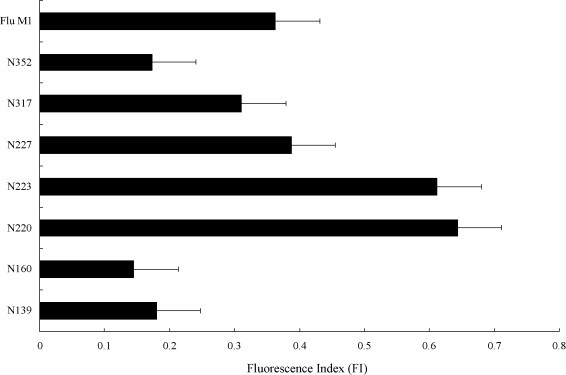
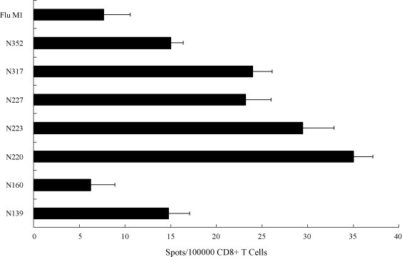
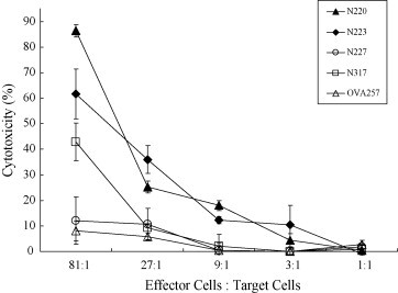
The severe acute respiratory syndrome coronavirus nucleocapsid protein (SARS-CoV N) is one of the major targets for SARS vaccine due to its high potency in triggering immune responses. In this study, we have identified a novel HLA-A*0201 restricted epitope, N220 (LALLLLDRL), of the SARS-CoV N-protein through bioinformatics analysis. The N-protein peptide N220 shows a high binding affinity towards human MHC class I in T2-cells, and is capable of activating cytotoxic T-cells in human peripheral blood mononuclear cells (PBMCs). The application of using the N220 peptide sequence with a single-chain-trimer (SCT) approach to produce a potential DNA vaccine candidate was investigated in HLA-A2.1K(b) transgenic mice. Cytotoxicity assay clearly showed that the T-cells obtained from the vaccinated animals were able to kill the N-protein expressing cells with a cytotoxicity level of 86% in an effector cells/target cells ratio of 81:1 one week after the last vaccination, which is significantly higher than other N-protein peptides previously described. The novel immunogenic N-protein peptide revealed in the present study provides valuable information for therapeutic SARS vaccine design.


Figures

Fig. 1
Fig. 1
A comparison of the binding affinity of the N-protein peptides towards the T2-cells. The N-protein peptides were incubated with the T2-cells in the presence of β2-microglobulin and the stabilized MHC-class I complexes were detected by an antibody (BB7.2) via flow cytometry. The relative amount of complexes formed is presented as the fluorescent index: [MFI (T2 + peptide)/MFI (T2 only)] − 1. The flu M1 peptide (GILGFVFTL) was used as a positive control. Results represent the mean ± S.D. (n = 5).
Fig. 2
Fig. 2
Activation of human T-cell in IFN-γ ELISPOT assay. CD8+ T-cells primed with N-protein peptides loaded dendritic cells were cultured with recombinant N protein-loaded autologous B-cells for 1 day and secretion of IFN-γ was measured by ELISPOT. Results represent the mean ± S.D. (n = 2) and the T-cells primed with an irrelevant flu peptide was used as a negative control.
Fig. 3
Fig. 3
Structure of the N-protein peptide with a single-chain-trimer gene. Different N-protein peptides and the OVA peptide were linked to the N-terminus of a fusion gene containing the human β2-microglobulin, human HLA-A2.1 α-1 domain, human HLA-A2.1 α-2 domain and mouse H2-Kb α-3 domain to construct the MHK gene. Linkers were inserted in between the N-protein peptide and the human β2-microglobulin, and the human β2-microglobulin and the human HLA-A2.1 α-1 domain, respectively.
Fig. 4
Fig. 4
Expression of the SARS-CoV N and the HPV16 E6-E7 genes in the N/E6E7/A2.1Kb cell line. After transduction of the N-protein gene and the HPV16 E6-E7 gene into the lung fibroblast cells of the HLA-A2.1Kb transgenic mice, total RNA of the selected cell clone was extracted. RT-PCR reaction was performed using oligo-dT primer in the first strand DNA synthesis followed by gene specific primers in the PCR reaction. From left to right, the first lane is the GADPH gene product, the second lane is the N gene product, and the third lane is E6-E7 gene product.
Fig. 5
Fig. 5
Cytotoxicity level of the spleen T-cells against the N/E6E7/A2.1Kb cells after DNA vaccination. T-cells of the spleen were harvested 1 week after the last vaccination and cytotoxicity against the N-protein expressing cells was compared based on the LDH release. The x-axis indicates the different ratios of the effector cells (splenocytes) to the target cells (N/E6E7/A2.1Kb). The y-axis indicates the percentage of cytotoxicity. Four groups of mice were vaccinated with the N-protein peptide plasmids, N220MHKpVAX1 (▴); N223MHKpVAX1 (♦); N227MHKpVAX1 (○); and N317MHKpVAX1 (□). Mice vaccinated with an irrelevant plasmid, OVAMHKpVAX1 (▵), was used as a negative control. The cytotoxicity level was calculated as previously described. The differences in cytotoxicity level between all peptides with various effector cells: target cells ratios were calculated with t-test (P < 0.05).

Similar articles

Cited by

References

    1. Chan P.K., Tang J.W., Hui D.S. SARS: clinical presentation, transmission, pathogenesis and treatment options. Clin Sci (Lond) 2006;110:193–204. - PubMed
    1. Ng W.F., To K.F., Lam W.W., Ng T.K., Lee K.C. The comparative pathology of severe acute respiratory syndrome and avian influenza A subtype H5N1—a review. Hum Pathol. 2006;37:381–390. - PMC - PubMed
    1. Poon L.L., Guan Y., Nicholls J.M., Yuen K.Y., Peiris J.S. The aetiology, origins, and diagnosis of severe acute respiratory syndrome. Lancet Infect Dis. 2004;4:663–671. - PMC - PubMed
    1. Tan Y.J., Lim S.G., Hong W. Characterization of viral proteins encoded by the SARS-coronavirus genome. Antiviral Res. 2005;65:69–78. - PMC - PubMed
    1. Ackerman A.L., Cresswell P. Cellular mechanisms governing cross-presentation of exogenous antigen. Nat Immunol. 2004;5:678–684. - PubMed

Publication types

MeSH terms