

Androgen and estrogen receptor-mediated mechanisms of testosterone action in male rat pelvic autonomic ganglia
- PMID: 17629410
- PMCID: PMC2012365
- DOI: 10.1016/j.neuroscience.2007.05.043
Androgen and estrogen receptor-mediated mechanisms of testosterone action in male rat pelvic autonomic ganglia
Abstract
Although male reproductive function is primarily androgen dependent, many studies suggest that estrogens have direct actions on the male reproductive organs. Pelvic autonomic neurons provide the motor control of the internal reproductive organs and the penis and various properties of these neurons are affected by endogenous androgens. However, the possible role of estrogens at this site has not been examined. Here we have investigated the significance of estrogens produced by aromatization of testosterone (T) in the physiological actions of androgens on adult male rat pelvic ganglion neurons. Reverse transcriptase polymerase chain reaction (RT-PCR) studies showed that aromatase and both estrogen receptors (ERalpha and ERbeta) are expressed in these ganglia. Western blotting also showed that aromatase is expressed in male pelvic ganglia. Using immunohistochemical visualization, ERalpha was predominantly expressed by nitric oxide synthase (NOS)-positive parasympathetic pelvic ganglion neurons. In vivo studies showed that the decrease in pelvic ganglion soma size caused by gonadectomy could be prevented by administration of T or dihydrotestosterone (DHT), but not 17beta-estradiol (E2), showing that this maintenance action of testosterone is mediated entirely by androgenic mechanisms. However, in vitro studies of cultured pelvic ganglion neurons revealed that T, DHT and E each stimulated the growth of longer and more complex neurites in both noradrenergic and cholinergic NOS-expressing neurons. The effects of T were attenuated by either androgen or estrogen receptor antagonists, or by inhibition of aromatase. Together these studies demonstrate that estrogens are likely to be synthesized in the male pelvic ganglia, produced from T by local aromatase. The effects of androgens on axonal growth are likely to be at least partly mediated by estrogenic mechanisms, which may be important for understanding disease-, aging- and injury-induced plasticity in this part of the nervous system.
Figures
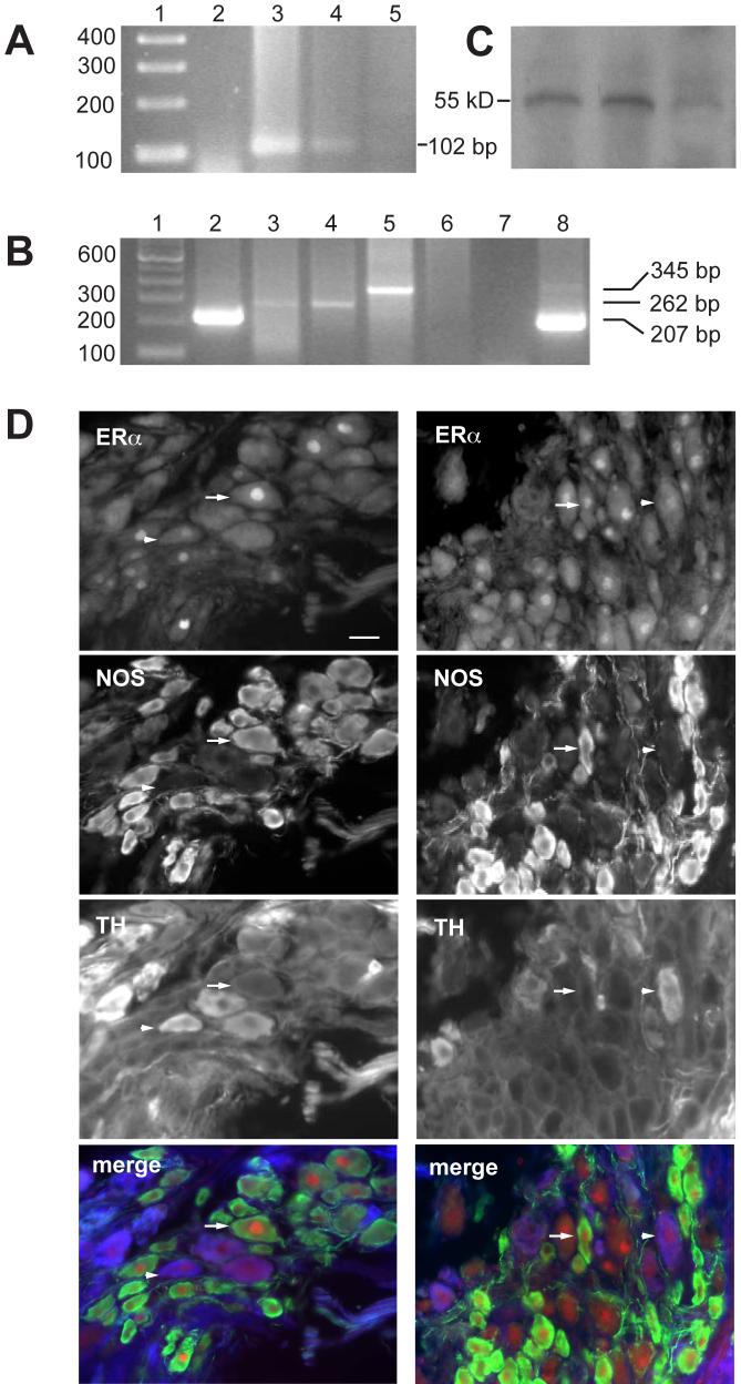
Aromatase RT-PCR yielded a 102 bp band in prostate and pelvic ganglion but not skeletal muscle; lane 1, 100 bp ladder; 2, blank; 3, prostate; 4, pelvic ganglion; 5, skeletal muscle.
ER RT-PCR yielded a 345 bp band for ERα and a 262 bp for ERβ in pelvic ganglion but only the ERβ transcript was present in prostate; lane 1, 100 bp ladder; 2, pelvic ganglion GAPDH (207 bp); 3, pelvic ganglion ERβ (262̣ bp); 4, prostate ERβ; 5, pelvic ganglion ERα(345 bp); 6, prostate ERα; 7 blank, ERα primers; 8, prostate GAPDH. For all primer sets, experiments were repeated twice.
Aromatase protein was identified in extracts of pelvic ganglia from three rats, each showing a single band at ∼55 kD.
Triple staining immunofluorescence showing examples of ERα expression in noradrenergic (TH-positive) and nitrergic (NOS-positive) pelvic ganglion neurons. Arrows show examples of ERα expression in NOS-positive, TH-negative neurons, and arrowheads show examples of weaker ERα expression in TH-positive, NOS-negative neurons. In merged colour images, ERα is red, NOS is green and TH is blue. Calibration in D represents 20 μm.
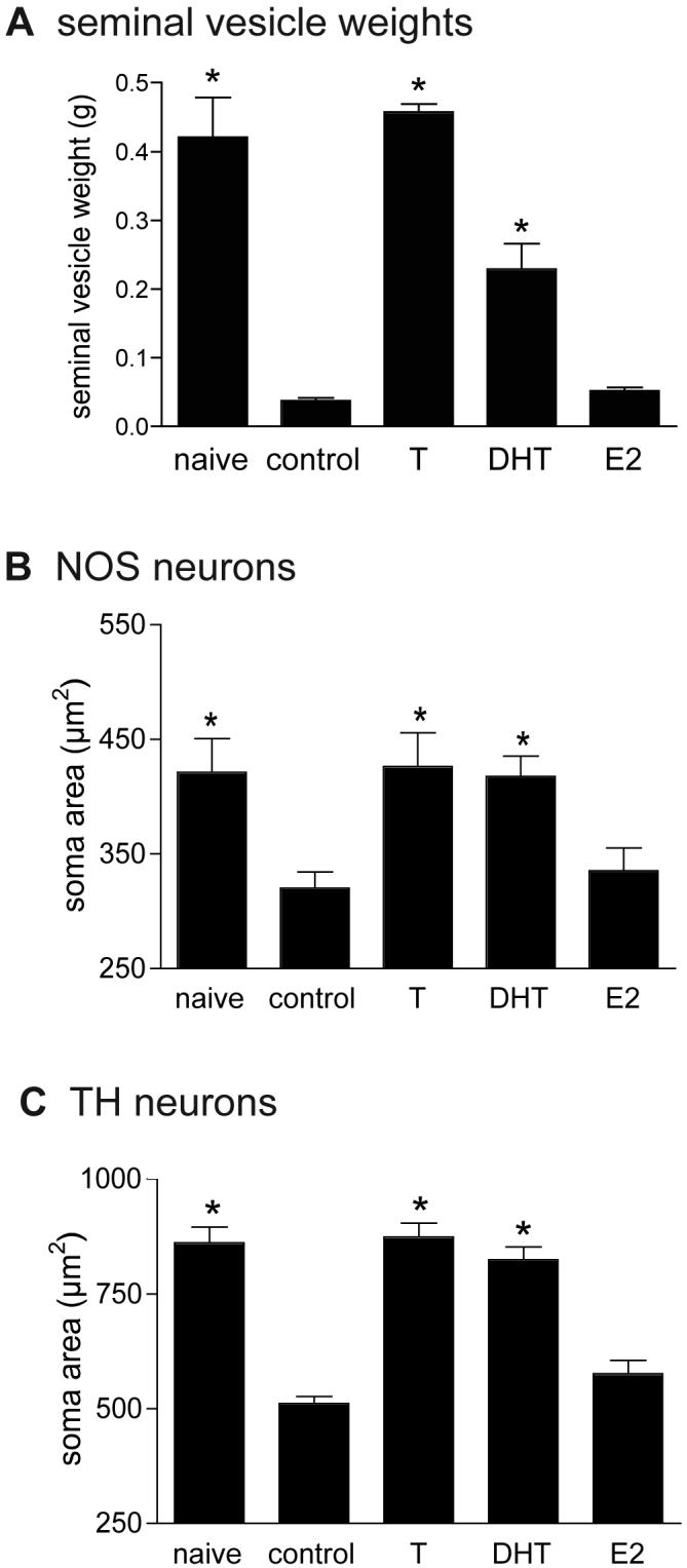
Seminal vesicles (mean of paired seminal vesicles in each animal) were much smaller after gonadectomy (n=5, “control”) compared to naïve intact animals (n=4) and this effect was completely prevented by testosterone (T, n=6), partly prevented by dihydrotestosterone (DHT, n=5) but unchanged by estradiol (E2, n=6). *, significantly different from control and from E2, P<0.001; not indicated, but DHT vs. naïve or T, P<0.001.
Size of NOS-immunoreactive somata decreased after gonadectomy (“control”) and this effect was completely prevented by T or DHT but not by E2. Data is from the same animals as shown in A. *, significantly different from control and from E2, P<0.05.
Size of TH-immunoreactive somata decreased after gonadectomy (“control”) and this effect was completely prevented by T or DHT but not by E2. Data is from the same animals as shown in A. *, significantly different from control and from E2, P<0.001.
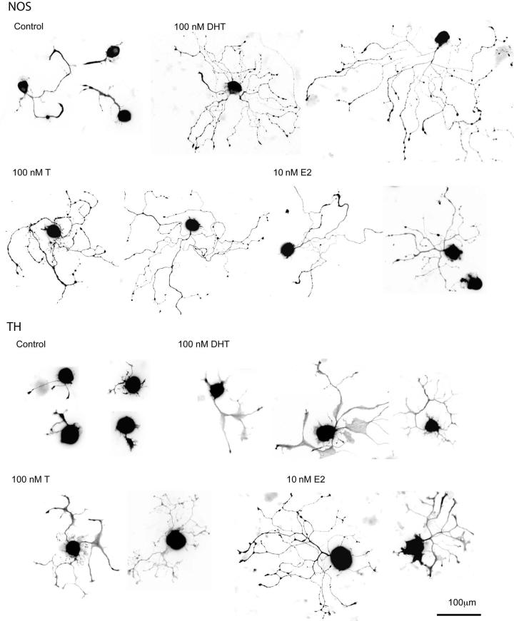
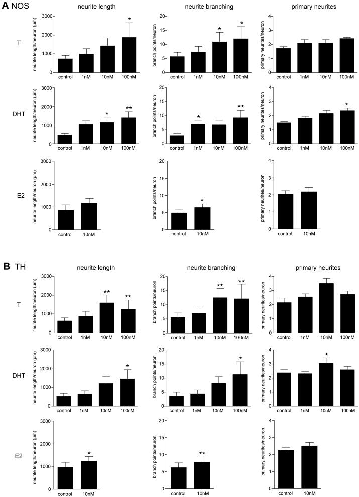
In NOS neurons, all three steroids increased neurite branching, both androgens increased neurite length and only DHT increased primary neurite number.
In TH neurons, all three steroids increased neurite length and branching but only DHT increased primary neurite number.
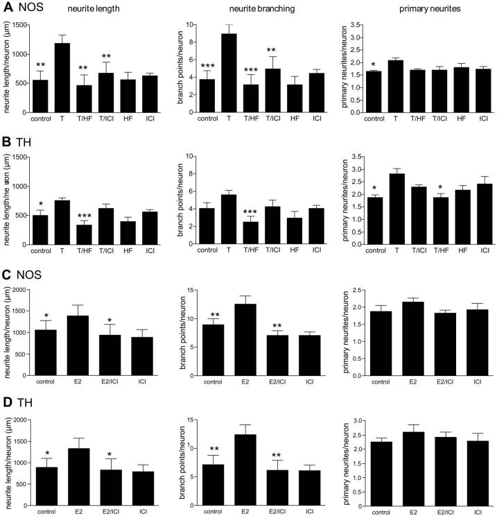
In NOS neurons, T (10 nM) caused an increase in neurite length (P<0.01), branching (P<0.001) and number (P<0.05) compared with untreated controls. The effects on length and branching were blocked by the AR blocker, hydroxyflutamide (HF, 10μM; T vs. T/HF, P<0.01 and P<0.001 for length and branching respectively) and by the ER blocker, ICI182780 (ICI, 10μM; T vs. T/ICI, P<0.01 for both parameters). The modest effects of T on neurite number appeared to be reduced by each blocker but neither effect reached statistical significance. Groups significantly different from T are indicated, *P<0.05, **P<0.01, ***P<0.001.
In TH neurons, T (10 nM) caused an increase in neurite length (P<0.05), and number (P<0.05) compared with untreated controls. A small effect of T on neurite branching appeared to occur but this was not statistically significant compared with controls; however, an effect of T on this parameter was indicated by the difference between T and T/HF groups (P<0.001). The effects on length and number were blocked by the AR inhibitor, hydroxyflutamide (HF, 10μM; T vs. T/HF, P<0.001 for length and P<0.05 for number). The ER blocker, ICI182780 (ICI, 10μM) had no significant effect on the T-induced increase in neurite length and branching. Groups significantly different from T are indicated, *P<0.05, ***P<0.001.
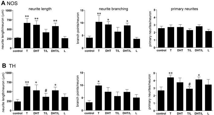
In NOS neurons, both T and DHT (each 10nM) stimulated neurite length and branching. The aromatase inhibitor letrozole (L, 100 nM) appeared to partially reduce the effects of testosterone (10 nM) on neurite length and branching but T/Lwas not significantly different to either control or T alone. Letrozole had no effect on the response to DHT (10 nM). In this group of experiments, androgens had weak or no effects on the number of primary neurites, but it should be noted that the controls showed a relatively high number of primary neurites compared with experiments shown in Figure 5. Groups significantly different from control are indicated, * P< 0.05, ** P < 0.01.
In TH neurons, the effects of T on neurite length and number of primary neurites were abolished by letrozole (L, 100 nM) (T vs T/L, P < 0.05), whereas the effect on neurite branching appeared to be only slightly reduced, i.e. T/L not significantly different to either control or T alone. The effects of DHT were unchanged by letrozole. Groups significantly different from control are indicated, * P< 0.05, ** P < 0.01. Groups significantly different from T are indicated, # P < 0.05.
Similar articles
-
Testosterone and nerve growth factor have distinct but interacting effects on structure and neurotransmitter expression of adult pelvic ganglion cells in vitro.Neuroscience. 2001;108(2):331-40. doi: 10.1016/s0306-4522(01)00420-1. Neuroscience. 2001. PMID: 11734365
-
Disruption of classical estrogenic targets in brown trout primary hepatocytes by the model androgens testosterone and dihydrotestosterone.Aquat Toxicol. 2020 Oct;227:105586. doi: 10.1016/j.aquatox.2020.105586. Epub 2020 Aug 15. Aquat Toxicol. 2020. PMID: 32882451
-
Mechanism of action of bolandiol (19-nortestosterone-3beta,17beta-diol), a unique anabolic steroid with androgenic, estrogenic, and progestational activities.J Steroid Biochem Mol Biol. 2010 Feb 15;118(3):151-61. doi: 10.1016/j.jsbmb.2009.11.008. Epub 2009 Nov 24. J Steroid Biochem Mol Biol. 2010. PMID: 19941958 Free PMC article.
-
Effects of testosterone and its metabolites on aromatase-immunoreactive cells in the quail brain: relationship with the activation of male reproductive behavior.J Steroid Biochem Mol Biol. 1996 Jan;56(1-6 Spec No):185-200. doi: 10.1016/0960-0760(95)00236-7. J Steroid Biochem Mol Biol. 1996. PMID: 8603040 Review.
-
Androgens and bone.Endocr Rev. 2004 Jun;25(3):389-425. doi: 10.1210/er.2003-0003. Endocr Rev. 2004. PMID: 15180950 Review.
Cited by
-
Neurophysiological control of urinary bladder storage and voiding-functional changes through development and pathology.Pediatr Nephrol. 2021 May;36(5):1041-1052. doi: 10.1007/s00467-020-04594-4. Epub 2020 May 15. Pediatr Nephrol. 2021. PMID: 32415328 Review.
-
The Effect of Castration on Peripheral Autonomic Neurons Supplying Mammalian Male Genitourinary System.Int J Mol Sci. 2021 Jul 16;22(14):7632. doi: 10.3390/ijms22147632. Int J Mol Sci. 2021. PMID: 34299251 Free PMC article. Review.
-
The Anatomy, Histology, and Function of the Major Pelvic Ganglion.Animals (Basel). 2024 Sep 4;14(17):2570. doi: 10.3390/ani14172570. Animals (Basel). 2024. PMID: 39272355 Free PMC article. Review.
-
Exposure to Organophosphate and Neonicotinoid Insecticides and Its Association with Steroid Hormones among Male Reproductive-Age Farmworkers in Northern Thailand.Int J Environ Res Public Health. 2021 May 24;18(11):5599. doi: 10.3390/ijerph18115599. Int J Environ Res Public Health. 2021. PMID: 34073889 Free PMC article.
-
Expression of Glial Cell Line-Derived Neurotrophic Factor (GDNF) and the GDNF Family Receptor Alpha Subunit 1 in the Paravaginal Ganglia of Nulliparous and Primiparous Rabbits.Int Neurourol J. 2018 Jan;22(Suppl 1):S23-33. doi: 10.5213/inj.1834974.487. Epub 2018 Jan 31. Int Neurourol J. 2018. PMID: 29385786 Free PMC article.
References
-
- Alm P, Uvelius B, Ekstrom J, Holmqvist B, Larsson B, Andersson KE. Nitric oxide synthase-containing neurons in rat parasympathetic, sympathetic and sensory ganglia: a comparative study. Histochem J. 1995;27:819–831. - PubMed
-
- Bianco JJ, Handelsman DJ, Pedersen JS, Risbridger GP. Direct response of the murine prostate gland and seminal vesicles to estradiol. Endocrinology. 2002;143:4922–4933. - PubMed
-
- Blacklock AD, Cauveren JA, Smith PG. Estrogen selectively increases sensory nociceptor innervation of arterioles in the female rat. Brain Res. 2004;1018:55–65. - PubMed
-
- Buchanan G, Birrell SN, Peters AA, Bianco-Miotto T, Ramsay K, Cops EJ, Yang M, Harris JM, Simila HA, Moore NL, Bentel JM, Ricciardelli C, Horsfall DJ, Butler LM, Tilley WD. Decreased androgen receptor levels and receptor function in breast cancer contribute to the failure of response to medroxyprogesterone acetate. Cancer Res. 2005;65:8487–8496. - PubMed
-
- Ceccatelli S, Lundberg JM, Zhang X, Aman K, Hökfelt T. Immunohistochemical demonstration of nitric oxide synthase in the peripheral autonomic nervous system. Brain Res. 1994;656:381–395. - PubMed
Publication types
MeSH terms
Substances
Grants and funding
LinkOut - more resources
Full Text Sources
