. 2007 Sep;90(3):397-406.
doi: 10.1016/j.ygeno.2007.05.004. Epub 2007 Jul 12.

Dissecting the human BDNF locus: bidirectional transcription, complex splicing, and multiple promoters

Affiliations

Dissecting the human BDNF locus: bidirectional transcription, complex splicing, and multiple promoters

Priit Pruunsild et al. Genomics. 2007 Sep.

Abstract

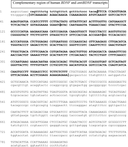
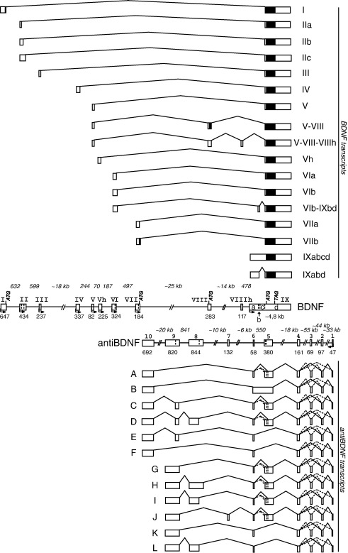
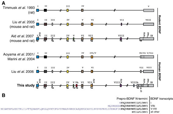
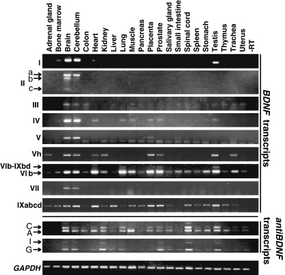
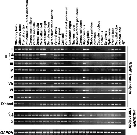
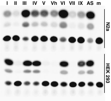
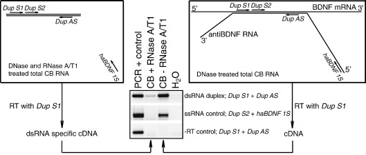
Brain-derived neurotrophic factor (BDNF), a member of the nerve growth factor family of neurotrophins, has central roles in the development, physiology, and pathology of the nervous system. We have elucidated the structure of the human BDNF gene, identified alternative transcripts, and studied their expression in adult human tissues and brain regions. In addition, the transcription initiation sites for human BDNF transcripts were determined and the activities of BDNF promoters were analyzed in transient overexpression assays. Our results show that the human BDNF gene has 11 exons and nine functional promoters that are used tissue and brain-region specifically. Furthermore, noncoding natural antisense RNAs that display complex splicing and expression patterns are transcribed in the BDNF gene locus from the antiBDNF gene (approved gene symbol BDNFOS). We show that BDNF and antiBDNF transcripts form dsRNA duplexes in the brain in vivo, suggesting an important role for antiBDNF in regulating BDNF expression in human.


Figures

Supplementary Figure 2
Fig. 1
Fig. 1
Structure and alternative transcripts of the human BDNF (top) and antiBDNF (bottom) genes. The structural organization of the exons and introns was determined by analyzing genomic and mRNA sequence data using bioinformatics, RT-PCR, and 5′ RACE. Exons are shown as boxes and introns as lines. Filled boxes and open boxes indicate the translated regions of the exons and the untranslated regions of the exons, respectively. The numbers below the exons and above the introns indicate their sizes. Exon and intron sizes are in base pairs, if not indicated otherwise. Arrows indicate the transcription start sites. ATG and TAG mark the positions of the translational start and stop codons, respectively. Vertical dashed lines indicate alternative splicing sites for the respective exons. BDNF exon IX is divided into regions “a”, “b”, “c”, and “d” as indicated in the box marking the position of exon IX. BDNF transcript names relate to the upstream exons used in front of the major 3′ exon IXd. “A”–“L” mark antiBDNF transcripts. Solid lines connecting the exons of transcripts represent the major splicing patterns of exons. Dashed lines connecting the exons of transcripts represent the minor splicing patterns of antiBDNF. Exon numbers are shown in Roman numerals for the BDNF gene and in Arabic numerals for the antiBDNF gene.
Fig. 2
Fig. 2
(A) Comparison of the human and rodent BDNF gene structures proposed by different studies. Structures presented are according to Timmusk et al. , Liu et al. , Aid et al. , Aoyama et al. , Marini et al. , and Liu et al. and the human BDNF gene structure determined in this study. Exons are shown as boxes and introns as lines. The identical human exons and the rodent exons homologous to the human exons are shown in the same color in all the structures. Novel exons determined by this study are in green (V), red (Vh), purple (VIII), and light blue (VIIIh). (B) Amino acid sequences of different potential prepro-BDNF N-termini. Amino acids encoded by exon IX are in black and sequences encoded by alternative 5′ exons are in blue. The transcripts encoding the respective N-termini of BDNF are listed adjacent to the N-terminal sequences.
Fig. 3
Fig. 3
Semiquantitative analysis of human BDNF, antiBDNF, and control GAPDH mRNA expression in adult human tissues by RT-PCR. Roman numerals on the left indicate the detected BDNF transcripts and the 5′ exon-specific primers used in combination with an antisense primer located in the BDNF coding region in exon IXd (Supplementary Table 1). A, C, G, and I refer to the respective antiBDNF transcripts shown in Fig. 1.
Fig. 4
Fig. 4
Semiquantitative analysis of human BDNF, antiBDNF, and control GAPDH mRNA expression by RT-PCR in different human brain regions. Roman numerals on the left indicate the detected BDNF transcripts and the 5′-exon-specific primers used in combination with an antisense primer located in the BDNF coding region in exon IXd (Supplementary Table 1). A, C, and G refer to the respective antiBDNF transcripts shown in Fig. 1.
Fig. 5
Fig. 5
Analyses of BDNF and antiBDNF promoter activities in HEK293T and N2a cells. The relative activities of the 5′ flanking regions of BDNF exons I, II, III, IV, V, Vh, VI, VII, and IXabcd and antiBDNF to promote CAT expression are shown. The promoter regions cloned in front of the CAT gene are shown in Supplementary Fig. 1. Note that the activities of BDNF promoters II, V, Vh, and VII in HEK239T cells were detectable using longer reaction times. m, mock-transfected cells, negative control.
Fig. 6
Fig. 6
BDNF and antiBDNF transcripts form dsRNA duplexes in the human brain in vivo. A schematic representation of the RNA duplex detection assay is shown. Briefly, total human cerebellar RNA was DNase treated. This RNA was divided into two—one half was treated with RNase A/T1 (box on the left) and the other was used as a − RNase control (box on the right). Both RNAs were reverse transcribed (RT) with a BDNF/antiBDNF complementary region-specific primer, Dup_S1. Subsequently these cDNAs were used as templates in PCR to detect BDNF/antiBDNF duplex with primers Dup_S1 and Dup_AS. ssRNA contamination control reaction was conducted with primers Dup_S2 and haBDNF_S1. The − RT reaction was used for detection of genomic DNA contamination using primers Dup_S1 and Dup_AS. Lines indicate RNAs, double line marks the complementary region of BDNF exon IXd and antiBDNF exon 5. Primer positions are indicated with arrows parallel with the lines and primer names are in italic. Human hippocampal cDNA synthesized using an oligo(dT) primer (PCR + control) was used as positive control for all the reactions.

References

    1. Binder D.K., Scharfman H.E. Brain-derived neurotrophic factor. Growth Factors. 2004;22:123–131. - PMC - PubMed
    1. Bibel M., Barde Y.A. Neurotrophins: key regulators of cell fate and cell shape in the vertebrate nervous system. Genes Dev. 2000;14:2919–2937. - PubMed
    1. Pezet S., McMahon S.B. Neurotrophins: mediators and modulators of pain. Annu. Rev. Neurosci. 2006;29:507–538. - PubMed
    1. Alsina B., Vu T., Cohen-Cory S. Visualizing synapse formation in arborizing optic axons in vivo: dynamics and modulation by BDNF. Nat. Neurosci. 2001;4:1093–1101. - PubMed
    1. Korte M., Carroll P., Wolf E., Brem G., Thoenen H., Bonhoeffer T. Hippocampal long-term potentiation is impaired in mice lacking brain-derived neurotrophic factor. Proc. Natl. Acad. Sci. USA. 1995;92:8856–8860. - PMC - PubMed

Publication types

Substances

Associated data