

Compensational regulation of bHLH transcription factors in the postnatal development of BETA2/NeuroD1-null retina
- PMID: 17629466
- PMCID: PMC4300853
- DOI: 10.1016/j.mod.2007.06.001
Compensational regulation of bHLH transcription factors in the postnatal development of BETA2/NeuroD1-null retina
Abstract
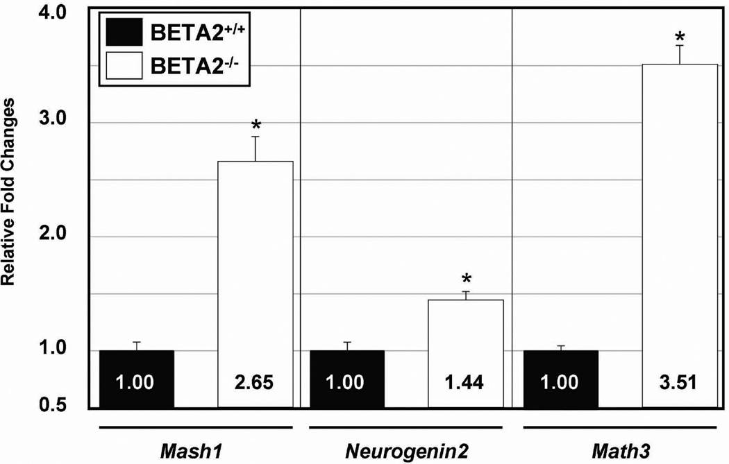
The bHLH transcriptional factor BETA2/NeuroD1 is essential for the survival of photoreceptor cells in the retina. Although this gene is expressed throughout the retina, BETA2/NeuroD1 knockout mice show photoreceptor cell degeneration only in the outer nuclear layer of the retina; other retinal neurons are not affected. Previous studies on retina explants lacking three bHLH genes revealed that retinal neurons in the inner nuclear layer require multiple bHLH genes for their differentiation and survival. However, single- or double-gene mutations show no or a lesser degree of abnormalities during eye development, likely because of compensation or cooperative regulation among those genes. Because not all null mice survive until the retina is fully organized, no direct evidence of this concept has been reported. To understand the regulatory mechanisms between bHLH factors in retinal development, we performed a detailed analysis of BETA2/NeuroD1 knockout mice. BETA2/NeuroD1 was expressed in all 3 layers of the mouse retina, including all major types of neurons. In addition, a null mutation of BETA2/NeuroD1 resulted in up-regulation of other bHLH genes, Mash1, Neurogenin2, and Math3, in the inner nuclear layer. Our data suggest that compensatory and cross regulatory mechanisms exist among the bHLH factors during retinal development.
Figures


References
-
- Acharya HR, Dooley CM, Thoreson WB, Ahmad I. cDNA cloning and expression analysis of NeuroD mRNA in human retina. Biochem. Biophys. Res. Commun. 1997;233:459–463. - PubMed
-
- Akagi T, Inoue T, Miyoshi G, Bessho Y, Takahashi M, Lee JE, Guillemot F, Kageyama R. Requirement of multiple basic helix-loop-helix genes for retinal neuronal subtype specification. J Biol Chem. 2004;279:28492–28498. - PubMed
-
- Brown NL, Kanekar S, Vetter ML, Tucker PK, Gemza DL, Glaser T. Math5 encodes a murine basic helix-loop-helix transcription factor expressed during early stages of retinal neurogenesis. Development. 1998;125:4821–4833. - PubMed
-
- Cepko CL. The roles of intrinsic and extrinsic cues and bHLH genes in the determination of retinal cell fates. Curr Opin Neurobiol. 1999;9:37–46. - PubMed
Publication types
MeSH terms
Substances
Grants and funding
LinkOut - more resources
Full Text Sources
Molecular Biology Databases
