Review
. 2007 Oct;19(5):488-96.
doi: 10.1016/j.coi.2007.05.004. Epub 2007 Jul 12.

Bcl-2-regulated apoptosis: mechanism and therapeutic potential

Affiliations
Review

Bcl-2-regulated apoptosis: mechanism and therapeutic potential

Jerry M Adams et al. Curr Opin Immunol. 2007 Oct.

Abstract

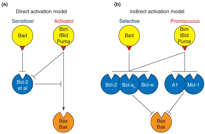
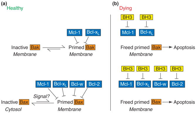
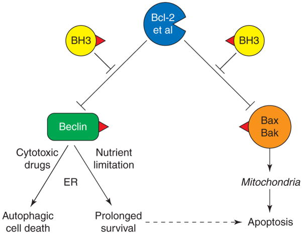
Apoptosis is essential for tissue homeostasis, particularly in the hematopoietic compartment, where its impairment can elicit neoplastic or autoimmune diseases. Whether stressed cells live or die is largely determined by interplay between opposing members of the Bcl-2 protein family. Bcl-2 and its closest homologs promote cell survival, but two other factions promote apoptosis. The BH3-only proteins sense and relay stress signals, but commitment to apoptosis requires Bax or Bak. The BH3-only proteins appear to activate Bax and Bak indirectly, by engaging and neutralizing their pro-survival relatives, which otherwise constrain Bax and Bak from permeabilizing mitochondria. The Bcl-2 family may also regulate autophagy and mitochondrial fission/fusion. Its pro-survival members are attractive therapeutic targets in cancer and perhaps autoimmunity and viral infections.


Figures

Figure 1
Figure 1
Two models for how BH3-only proteins activate Bax and Bak. (A) In the direct activation model [11], the indicated activator BH3-only proteins, via their BH3 domain (red triangle), directly engage Bax and Bak and activate them, whereas sensitizer BH3-only proteins (e.g. Bad or Noxa), which can only bind the pro-survival proteins, serve only to displace activators from the pro-survival proteins. (B) In the indirect activation model [10**,19**,20**] the BH3-only proteins only bind the pro-survival proteins. Because the promiscuous binders (Bim, tBid, Puma) can neutralise all pro-survival proteins, each can readily trigger Bax/Bak activation, whereas any selective binder (eg. Bad) must be co-expressed with a complementary binder (e.g. Noxa) to do so.
Figure 2
Figure 2
Model for constraint of Bak (top) and Bax (bottom) by pro-survival family members [19**,20**,21]. (A) In unperturbed cells or early in apoptotic signalling, a small proportion of Bak and Bax molecules is proposed to acquire a ‘primed’ conformation in which their BH3 domains are accessible but engaged by the indicated pro-survival proteins (and perhaps also A1). The priming might require an independent undetermined signal, particularly to recruit Bax from the cytosol. (B) In apoptotic cells, once BH3-only proteins (‘BH3’), or a BH3 mimic, have engaged the appropriate pro-survival proteins, the freed primed Bak or Bax can nucleate the oligomerization thought to permeabilize the outer mitochondrial membrane and thereby commit the cell to apoptosis. The pro-survival proteins also appear to have a mode of constraint that does not rely upon association via the Bax/Bak BH3 domain (see text).
Figure 3
Figure 3
Models for resistance/sensitivity of tumor cells to a selective BH3 mimetic like ABT-737 (red arrowhead) that binds tightly to Bcl-xL, Bcl-2 and Bcl-w but not to Mcl-1 or A1. (A) High Mcl-1 (dark blue) renders the cell refractory, because Mcl-1 can capture the Bak released from Bcl-xL by the drug. (B) In cells with little Mcl-1 (light blue) (or A1), the freed Bak can initiate apoptosis. (C) The high Bcl-2 (or Bcl-xL) found in many tumors can sequester a high level of Bim [44] and prevent Bim from inducing apoptosis. Paradoxically, this can actually enhance sensitivity to ABT-737, because the Bim released by ABT-737 from Bcl-2 (or Bcl-xL) can inactivate the ABT-737-insensitive Mcl-1 (or A1), freeing Bak to drive apoptosis.
Figure 4
Figure 4
Potential coupling of apoptosis and autophagy via Beclin 1 and Bcl-2 homologs. The model assumes that, in a favourable environment, pro-survival proteins such as Bcl-2 and Bcl-xL prevent Beclin 1 from inducing autophagy by sequestering it, seemingly on the ER [52**,53]. If so, the engagement of the pro-survival proteins by BH3-only proteins (or BH3 mimics) should trigger autophagy as well as apoptosis. In settings such as nutrient limitation or cytokine deprivation, autophagy temporarily prolongs survival by ensuring adequate ATP levels [49*,52**], but in response to cytotoxic drugs, including some used in chemotherapy, it appears instead to promote autophagic cell death [55**]. How this dichotomy is controlled is not known.

Similar articles

Cited by

References

    1. Strasser A. The role of BH3-only proteins in the immune system. Nat Rev Immunol. 2005;5:189–200. This recent review clearly outlines the physiological roles of the Bcl-2 family and apoptosis, particularly in the immune system. - PubMed
    1. Adams JM. Ways of dying: multiple pathways to apoptosis. Genes Dev. 2003;17:2481–2495. - PubMed
    1. Shi Y. Mechanical aspects of apoptosome assembly. Curr Opin Cell Biol. 2006;18:677–684. - PubMed
    1. Adams JM, Cory S. The Bcl-2 apoptotic switch in cancer development and therapy. Oncogene. 2007;26:1324–1337. This recent review focuses on the mechanisms by which the Bcl-2 family control apoptosis and their impact on cancer development and therapy. - PMC - PubMed
    1. Hinds MG, Day CL. Regulation of apoptosis: uncovering the binding determinants. Curr Opin Struct Biol. 2005;15:690–699. - PubMed

Publication types

MeSH terms