

Enzymatic synthesis of glycosaminoglycan heparin
- PMID: 17629842
- PMCID: PMC4140616
- DOI: 10.1055/s-2007-982076
Enzymatic synthesis of glycosaminoglycan heparin
Abstract
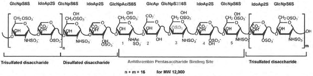
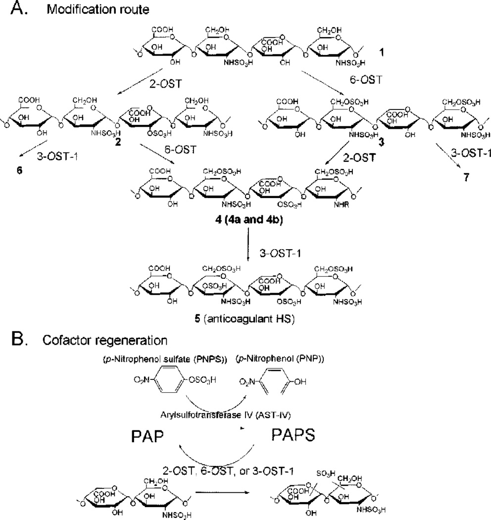
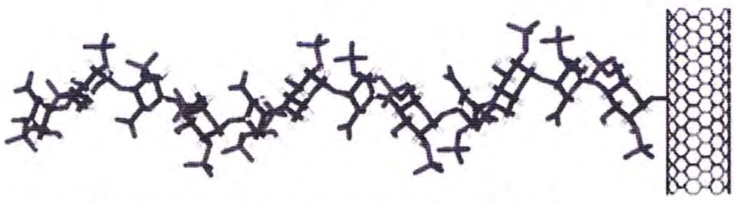
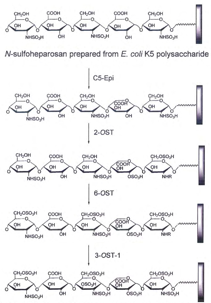
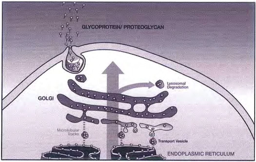
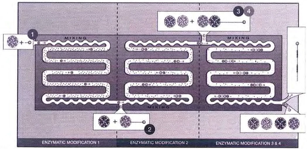
Heparin and its low molecular weight heparin derivatives, widely used as clinical anticoagulants, are acidic polysaccharide members of a family of biomacromolecules called glycosaminoglycans (GAGs). Heparin and the related heparan sulfate are biosynthesized in the Golgi apparatus of eukaryotic cells. Heparin is a polycomponent drug that currently is prepared for clinical use by extraction from animal tissues. A heparin pentasaccharide, fondaparinux, has also been prepared through chemical synthesis for use as a homogenous anticoagulant drug. Recent enabling technologies suggest that it may now be possible to synthesize heparin and its derivatives enzymatically. Moreover, new technologies including advances in synthetic carbohydrate synthesis, enzyme-based GAG synthesis, micro- and nano-display of GAGs, rapid on-line structural analysis, and microarray/microfluidic technologies might be applied to the enzymatic synthesis of heparins with defined structures and exhibiting selected activities. The advent of these new technologies also makes it possible to consider the construction of an artificial Golgi to increase our understanding of the cellular control of GAG biosyntheses in this organelle.
Figures
Similar articles
-
Chemoenzymatic Synthesis of Glycosaminoglycans.Acc Chem Res. 2020 Feb 18;53(2):335-346. doi: 10.1021/acs.accounts.9b00420. Epub 2019 Nov 12. Acc Chem Res. 2020. PMID: 31714740
-
Turkey intestine as a commercial source of heparin? Comparative structural studies of intestinal avian and mammalian glycosaminoglycans.Comp Biochem Physiol B Biochem Mol Biol. 2003 Jan;134(1):189-97. doi: 10.1016/s1096-4959(02)00250-6. Comp Biochem Physiol B Biochem Mol Biol. 2003. PMID: 12524047
-
Recent progress and applications in glycosaminoglycan and heparin research.Curr Opin Chem Biol. 2009 Dec;13(5-6):633-40. doi: 10.1016/j.cbpa.2009.08.017. Epub 2009 Sep 24. Curr Opin Chem Biol. 2009. PMID: 19781979 Free PMC article. Review.
-
Recent chemical and enzymatic approaches to the synthesis of glycosaminoglycan oligosaccharides.Curr Med Chem. 2003 Oct;10(19):1993-2031. doi: 10.2174/0929867033456891. Curr Med Chem. 2003. PMID: 12871100 Review.
-
Strategies in Synthesis of Heparin/Heparan Sulfate Oligosaccharides: 2000-Present.Adv Carbohydr Chem Biochem. 2021;80:121-164. doi: 10.1016/bs.accb.2021.11.003. Adv Carbohydr Chem Biochem. 2021. PMID: 34872655
Cited by
-
Fluorous supported modular synthesis of heparan sulfate oligosaccharides.Org Lett. 2013 Jan 18;15(2):342-5. doi: 10.1021/ol303270v. Epub 2013 Jan 8. Org Lett. 2013. PMID: 23293947 Free PMC article.
-
Platelet factor 4 polyanion immune complexes: heparin induced thrombocytopenia and vaccine-induced immune thrombotic thrombocytopenia.Thromb J. 2021 Sep 15;19(1):66. doi: 10.1186/s12959-021-00318-2. Thromb J. 2021. PMID: 34526009 Free PMC article. Review.
-
Quantification of heparan sulfate disaccharides using ion-pairing reversed-phase microflow high-performance liquid chromatography with electrospray ionization trap mass spectrometry.Anal Chem. 2009 Jun 1;81(11):4349-55. doi: 10.1021/ac9001707. Anal Chem. 2009. PMID: 19402671 Free PMC article.
-
Polysaccharide-modified synthetic polymeric biomaterials.Biopolymers. 2010;94(1):128-40. doi: 10.1002/bip.21334. Biopolymers. 2010. PMID: 20091875 Free PMC article. Review.
-
Characterization of heparin and severe acute respiratory syndrome-related coronavirus 2 (SARS-CoV-2) spike glycoprotein binding interactions.Antiviral Res. 2020 Sep;181:104873. doi: 10.1016/j.antiviral.2020.104873. Epub 2020 Jul 10. Antiviral Res. 2020. PMID: 32653452 Free PMC article.
References
-
- Linhardt RJ. Heparin: an important drug enters its seventh decade. Chem Ind. 1991;2:45–50.
-
- Linhardt RJ, Gunay NS. Production and chemical processing of low molecular weight heparins. Semin Thromb Hemost. 1999;25:5–14. - PubMed
-
- Casu B. Structure and biological activity of heparin. Adv Carbohydr Chem Biochem. 1985;43:51–134. - PubMed
-
- Fareed J, Jeske W, Hoppensteadt D, Clarizio R, Walgenga JM. Low molecular weight heparins: pharmacologic profile and product differentiation. Am J Cardiol. 1998;82:3L–10L. - PubMed
Publication types
MeSH terms
Substances
Grants and funding
LinkOut - more resources
Full Text Sources
Other Literature Sources
Medical
