

Early postnatal alcohol exposure reduced the size of vibrissal barrel field in rat somatosensory cortex (SI) but did not disrupt barrel field organization
- PMID: 17630086
- PMCID: PMC2435073
- DOI: 10.1016/j.alcohol.2007.04.005
Early postnatal alcohol exposure reduced the size of vibrissal barrel field in rat somatosensory cortex (SI) but did not disrupt barrel field organization
Abstract
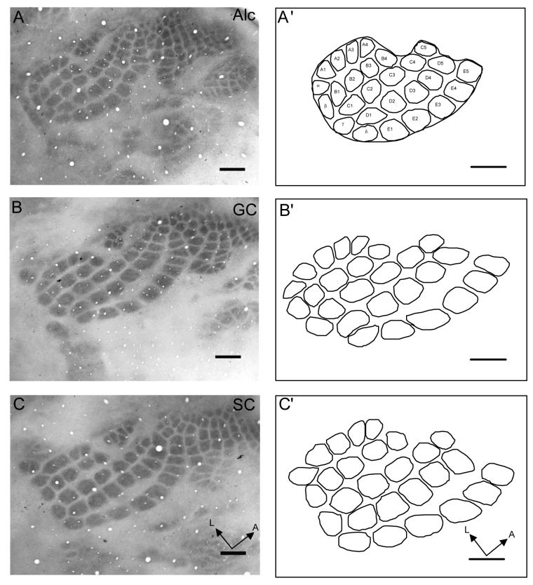
Prenatal alcohol exposure (PAE) has been shown to alter the somatosensory cortex in both human and animal studies. In rodents, PAE reduced the size, but not the pattern of the posteromedial barrel subfield (PMBSF) associated with the representation of the whiskers, in newborn, juvenile, and adult rats. However, the PMBSF is not present at birth, but rather first appears in the middle of the first postnatal week during the brain-growth spurt period. These findings raise questions whether early postnatal alcohol exposure might disrupt both barrel field pattern and size, questions that were investigated in the present study. Newborn Sprague-Dawley rats were assigned into alcohol (Alc), nutritional gastric control (GC), and suckle control (SC) groups on postnatal day 4 (P4). Rat pups in Alc and GC were artificially fed with alcohol and maltose-dextrin dissolved in milk, respectively, via an implant gastrostomy tube, from P4 to P9. Pups in the Alc group received alcohol (6.0 g/kg) in milk, while the GC controls received isocaloric equivalent maltose-dextrin dissolved in milk. Pups in the SC group remained with their mothers and breast fed throughout the experimental period. On P10, pups in each group were weighed, sacrificed, and their brains removed and weighed. Cortical hemispheres were separated, weighed, flattened, sectioned tangentially, stained with cytochrome oxidase, and PMBSF measured. The sizes of barrels and the interbarrel septal region within PMBSF, as well as body and brain weights were compared between the three groups. The sizes of PMSBF barrel and septal areas were significantly smaller (P<.01) in Alc group compared to controls, while the PMBSF barrel pattern remained unaltered. Body, whole-brain, forebrain, and hemisphere weights were significantly reduced (P<.01) in Alc pups compared to control groups. GC and SC groups did not differ significantly in all dependent variables, except body weight at P9 and P10 (P<.01). These results suggest that postnatal alcohol exposure, like prenatal exposure, significantly influenced the size of the barrel field, but not barrel field pattern formation, indicating that barrel field pattern formation consolidated prior to P4. These results are important for understanding sensorimotor deficits reported in children suffering from fetal alcohol spectrum disorder (FASD).
Figures
References
-
- Bayer SA, Altman J, Russo RJ, Zhang X. Timetables of neurogenesis in the human brain based on experimentally determined patterns in the rat. Neurotoxicology. 1993;14:83–144. - PubMed
-
- Bonthius DJ, Goodlett CR, West JR. Blood alcohol concentration and severity of microencephaly in neonatal rats depend on the pattern of alcohol administration. Alcohol. 1988;5:209–214. - PubMed
-
- Bonthius DJ, West JR. Blood alcohol concentration and microencephaly: a dose-response study in the neonatal rat. Teratology. 1988;37:223–231. - PubMed
-
- Bonthius DJ, West JR. Permanent neuronal deficits in rats exposed to alcohol during the brain growth spurt. Teratology. 1991;44:147–163. - PubMed
-
- Burd L, Cotsonas-Hassler TM, Martsolf JT, Kerbeshian J. Recognition and management of fetal alcohol syndrome. Neurotoxicol Teratol. 2003;25:681–688. - PubMed
Publication types
MeSH terms
Substances
Grants and funding
LinkOut - more resources
Full Text Sources
Miscellaneous
