. 2007 Jul;19(7):2278-92.
doi: 10.1105/tpc.107.052985. Epub 2007 Jul 13.

A unique beta1,3-galactosyltransferase is indispensable for the biosynthesis of N-glycans containing Lewis a structures in Arabidopsis thaliana

Affiliations

A unique beta1,3-galactosyltransferase is indispensable for the biosynthesis of N-glycans containing Lewis a structures in Arabidopsis thaliana

Richard Strasser et al. Plant Cell. 2007 Jul.

Abstract

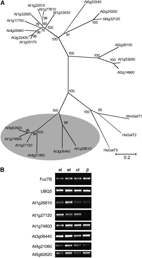
In plants, the only known outer-chain elongation of complex N-glycans is the formation of Lewis a [Fuc alpha1-4(Gal beta1-3)GlcNAc-R] structures. This process involves the sequential attachment of beta1,3-galactose and alpha1,4-fucose residues by beta1,3-galactosyltransferase and alpha1,4-fucosyltransferase. However, the exact mechanism underlying the formation of Lewis a epitopes in plants is poorly understood, largely because one of the involved enzymes, beta1,3-galactosyltransferase, has not yet been identified and characterized. Here, we report the identification of an Arabidopsis thaliana beta1,3-galactosyltransferase involved in the biosynthesis of the Lewis a epitope using an expression cloning strategy. Overexpression of various candidates led to the identification of a single gene (named GALACTOSYLTRANSFERASE1 [GALT1]) that increased the originally very low Lewis a epitope levels in planta. Recombinant GALT1 protein produced in insect cells was capable of transferring beta1,3-linked galactose residues to various N-glycan acceptor substrates, and subsequent treatment of the reaction products with alpha1,4-fucosyltransferase resulted in the generation of Lewis a structures. Furthermore, transgenic Arabidopsis plants lacking a functional GALT1 mRNA did not show any detectable amounts of Lewis a epitopes on endogenous glycoproteins. Taken together, our results demonstrate that GALT1 is both sufficient and essential for the addition of beta1,3-linked galactose residues to N-glycans and thus is required for the biosynthesis of Lewis a structures in Arabidopsis. Moreover, cell biological characterization of a transiently expressed GALT1-fluorescent protein fusion using confocal laser scanning microscopy revealed the exclusive location of GALT1 within the Golgi apparatus, which is in good agreement with the proposed physiological action of the enzyme.


Figures

Figure 1.
Figure 1.
Plant N-Glycan Structures. Characteristic plant N-glycan structures are shown. The paucimannosidic structure MMXF (for nomenclature, see www.proglycan.com and Supplemental Figure 5 online), which lacks terminal GlcNAc residues at the nonreducing ends, and the complex N-glycan GnGnXF are the major N-glycan species identified in plants. The Lea structure (FA)(FA)XF represents the most sophisticated complex plant N-glycan structure so far.
Figure 2.
Figure 2.
Presence of Lea Epitopes in Arabidopsis. Protein gel blot analysis and mass spectrometry of N-glycans were performed to detect the Lea carbohydrate epitope in individual Arabidopsis tissues. (A) Proteins were extracted from different organs, and 12 μg of total protein were subjected to SDS-PAGE under reducing conditions. Immunoblotting was performed using JIM84 antibodies. fl, flowers; pe, pedicels; si, siliques; st, stem; n, node; sa, shoot apex; cl, cauline leaves; jl, juvenile leaves; al, adult leaves, p, petiole; sl, senescent leaves; r, roots. (B) N-glycans isolated from wild-type stems were analyzed by MALDI-TOF MS. Two Lea peaks [(FA)GnXF/Gn(FA)XF and (FA)(FA)XF] were clearly detected.
Figure 3.
Figure 3.
Phylogenetic and Expression Analysis of Putative Arabidopsis GALTs. (A) Phylogenetic analysis of putative Arabidopsis GALT proteins was conducted using MEGA version 3.1. Amino acid sequences of the 20 pfam 01762-containing Arabidopsis proteins from CAZy glycosyltransferase family GT31 and three mammalian B3GALTs (Mm-B3GALT1, UniProt accession number O54904; Hs-B3GALT2, O43825; and Hs-B3GALT5, Q9BYG0) were aligned using ClustalW. The phylogenetic tree was constructed with the neighbor-joining method, and the bootstrap values were determined from 1000 trials. The percentages of bootstrap values for the respected branches are shown. The members of subfamily 1 are highlighted in gray. Bar = 20% divergence. (B) Expression analysis of GALT candidate genes by RT-PCR. RT-PCR was performed with specific primers for the respective genes from subfamily 1 or ubiquitin (UBQ5) and core α1,3-fucosyltransferase (FucTB) as controls (35 PCR cycles for all reactions). Siliques (si), stems (st), cauline leaves (cl), and juvenile leaves (jl) were analyzed. All RT-PCR reactions were performed twice with similar results.
Figure 4.
Figure 4.
Overexpression of GALT1 in Leaves. (A) Extracted leaf proteins from different transgenic lines (1 to 12) expressing 35S:GALT1 (10 μg) were subjected to SDS-PAGE under reducing conditions. Immunoblotting was performed using JIM84 antibody. Wild-type plants and 35S:Mm-B3GALT1 (Mm) plants were used as controls. (B) MALDI-TOF MS spectra of N-glycans extracted from wild-type and 35S:GALT1 (line 12) leaves. The peaks containing Lea structures are marked, and their masses are shown in the table.
Figure 5.
Figure 5.
GALT1 Amino Acid Sequence. The putative transmembrane domain (amino acids 6 to 23) predicted by HmmTOP_V2 (http://www.enzim.hu/hmmtop/index.html) is highlighted in gray, and putative N-glycosylation sites are underlined. The galactoside binding lectin domain (pfam 00337) is shown in italics, and the galactosyltransferase domain (pfam 01762) is shown in bold. The conserved DXD motif is shaded black, and the asterisk marks the beginning of the fragment that was expressed in insect cells as a soluble secreted form.
Figure 6.
Figure 6.
Recombinant GALT1 Modifies Glycopeptide and N-Glycan Substrates with β1,3-Linked Galactose Residues. (A) Immunoblot of purified recombinant GALT1, with (+) and without (−) PNGase F digestion. (B) Dabsylated GnGn glycopeptide (m/z = 2061) was incubated with purified recombinant GALT1 in the presence of the donor substrate UDP-galactose and analyzed by MALDI-TOF MS. In the bottom panel, the acceptor substrate (S) is shown as a control. Asterisks mark substrate fragmentation peaks. (C) GnGn-PA was incubated either with recombinant GALT1 or recombinant Mm-B3GALT1 and UDP-galactose. The reaction products of GALT1 coeluted with the Mm-B3GALT1 products. (D) Reaction products from the GALT1 assay shown in (C) were incubated with different β-galactosidases prior to rechromatography. X.m. galactosidase, β1,3-galactosidase from X. manihotis; A.o. galactosidase, A. oryzae β1,4-/β1,6-specific galactosidase.
Figure 7.
Figure 7.
Generation of the Lea Epitope on Glycopeptide and Glycoprotein Substrates. (A) Dabsylated GnGn-glycopeptide (m/z = 2061) was incubated with recombinant GALT1 in the presence of UDP-galactose and analyzed by MALDI-TOF MS (bottom panel). Subsequently, the product was incubated with recombinant FUT13 and GDP-fucose and analyzed by MALDI-TOF MS (top panel), which resulted in the detection of the (FA)(FA) peak (mass: 2677 D). Asterisks mark substrate and product fragmentation peaks. (B) GnGn-transferrin (GnGn-Tf) was incubated first with recombinant GALT1 and UDP-galactose and then with recombinant FUT13 and GDP-fucose. As controls the acceptor substrate (GnGn-Tf), the donor substrates (UDP-galactose and GDP-fucose), or the glycosyltransferases (GALT1 and FUT13) were omitted in different combinations. The samples were subjected to SDS-PAGE under reducing conditions, and after blotting, Lea epitopes were detected using JIM84 antibodies. Tf, transferrin; Tf*, β1,3-galactosylated contaminant present in the transferrin sample used in this experiment. Note that GALT1 itself serves also as a substrate for FUT13.
Figure 8.
Figure 8.
GALT1 Is Capable of in Vivo Autogalactosylation. (A) Recombinant GALT1 (5 μg) was incubated (16 h, 37°C) in 30 μL of 50 mM sodium citrate buffer, pH 4.5, either in the absence (lane 1) or presence of 42 milliunits (mU) of β1,3-galactosidase (lane 2) or 150 mU of β1,4-/β1,6-galactosidase (lane 3). Aliquots (2.5 μg GALT1) were then treated (16 h, 30°C) with recombinant FUT13 (5 μg) in 50 μL of 100 mM MES buffer, pH 7.0, containing 0.5 mM GDP-fucose. The samples were then subjected to immunoblotting analysis with antibodies to the Lea epitope (JIM84) or to the enterokinase cleavage site present in recombinant GALT1 (anti-EK). All samples contain a GALT1 fragment (GALT1*) that fails to react with anti-EK antibodies. (B) Recombinant GALT1 (5 μg) was excised from a Coomassie blue–stained SDS-PAGE gel and subjected to tryptic digestion and subsequent MS analysis to identify glycopeptides with terminal galactose residues. To distinguish between the isobaric structures Man4GnF and β1,3-galactosylated AMF/MAF, a control sample (right panel) was digested with X. manihotis β1,3-galactosidase (X.m.). This confirmed that the peak with the mass of 2672.1 D consists mainly of the β1,3-galactosylated structure AMF/MAF, with the β1,3-galactosidase–insensitive fraction probably representing Man4GnF. The relevant part of the mass spectrum for glycopeptide T279SSTSLQSNTSR290 is shown. The complete mass spectra of this and other GALT1 glycopeptides are included in Supplemental Figure 7 online.
Figure 9.
Figure 9.
GALT1 Uses GnGnXF as a Substrate. (A) The typical complex plant N-glycan substrate GnGnXF-PA was incubated with GALT1 and UDP-galactose to show that this β1,2-xylose and core α1,3-fucose containing N-glycan can act as an acceptor structure for GALT1. Three peaks were detected (GnA3XF-PA, A3GnXF-PA, and A3A3XF-PA), which coeluted with standards obtained by incubation of the substrate with Mm-B3GALT1. (B) The reaction products of the GALT1 assay were further modified by incubation with recombinant FUT13 and GDP-fucose. As shown by LC-MS analysis, this led to the formation of N-glycans carrying both β1,3-linked galactose and α1,4-linked fucose [i.e., (AF)GnXF/Gn(AF)XF and (AF)(AF)XF].
Figure 10.
Figure 10.
Analysis of RNAi and Knockout Plants. (A) The structure of the GALT1 gene and the position of the T-DNA insertion are shown. (B) PCR screening of the T-DNA knockout line (SAIL_170_A08) resulted in the identification of plants homozygous for the T-DNA insertion (galt1-1). Sequencing of the PCR products obtained with one gene-specific primer (At1g26810-1F) and one T-DNA–specific primer (LBsail2, T-DNA 1) or At1g26810-3R and LBsail2 (T-DNA 2) confirmed the insertion and revealed a deletion of 36 bp in intron 4 of GALT1. RT-PCR was performed on RNA extracted from galt1-1 plants with primers At1g26810-1F and At1g26810-2R. UBQ5 served as a control. (C) Schematic presentation of the GALT1 RNAi construct. (D) RT-PCR of a galt1RNAi line using RNA extracted from stems with GALT1-specific primers (AT1g26810-1F/-2R). FucTB served as a control. (E) Protein gel blot in which proteins were extracted from stems, and 10 μg were subjected to SDS-PAGE under reducing conditions. Detection was performed either with JIM84 or with anti-HRP antibodies, which recognize β1,2-xylose and core α1,3-fucose residues on N-glycans, as a loading control. While the Lea epitope is present in wild-type plants (lane 1) and in the 35S:GALT1 line (lane 3), it is not detectable in transgenic galt1-1 (lane 2) and galt1RNAi (lane 4) plants.
Figure 11.
Figure 11.
GALT1-GFP Accumulates in the Golgi. N. benthamiana leaf epidermal cells transiently expressing GALT1-GFP ([A] and [D]), ST-mRFP (B), and YFP-HDEL (E) and the corresponding overlays of the images ([C] and [F]). The punctate fluorescence of GALT1-GFP and its colocalization with ST-mRFP strongly indicates that GALT1-GFP accumulates in the Golgi, which is in good agreement with its proposed physiological action. Bars = 10 μm.

Similar articles

Cited by

References

    1. Ackerman, S., Liu, L., Kwatia, M., Savage, M., Leonidas, D., Swaminathan, G., and Acharya, K. (2002). Charcot-Leyden crystal protein (galectin-10) is not a dual function galectin with lysophospholipase activity but binds a lysophospholipase inhibitor in a novel structural fashion. J. Biol. Chem. 277 14859–14868. - PubMed
    1. Altmann, F., Schweiszer, S., and Weber, C. (1995). Kinetic comparison of peptide: N-glycosidases F and A reveals several differences in substrate specificity. Glycoconj. J. 12 84–93. - PubMed
    1. Amado, M., Almeida, R., Carneiro, F., Levery, S., Holmes, E., Nomoto, M., Hollingsworth, M., Hassan, H., Schwientek, T., Nielsen, P., Bennett, E., and Clausen, H. (1998). A family of human beta3-galactosyltransferases. Characterization of four members of a UDP-galactose:beta-N-acetyl-glucosamine/beta-nacetyl-galactosamine beta-1,3-galactosyltransferase family. J. Biol. Chem. 273 12770–12778. - PubMed
    1. Bakker, H., Bardor, M., Molthoff, J., Gomord, V., Elbers, I., Stevens, L., Jordi, W., Lommen, A., Faye, L., Lerouge, P., and Bosch, D. (2001. b). Galactose-extended glycans of antibodies produced by transgenic plants. Proc. Natl. Acad. Sci. USA 98 2899–2904. - PMC - PubMed
    1. Bakker, H., Schijlen, E., de Vries, T., Schiphorst, W., Jordi, W., Lommen, A., Bosch, D., and van Die, I. (2001. a). Plant members of the alpha1-3/4-fucosyltransferase gene family encode an alpha1-4-fucosyltransferase, potentially involved in Lewis(a) biosynthesis, and two core alpha1-3-fucosyltransferases. FEBS Lett. 507 307–312. - PubMed

Publication types

MeSH terms

Associated data