. 2007 Sep;6(9):1646-55.
doi: 10.1128/EC.00177-07. Epub 2007 Jul 13.

EhLimA, a novel LIM protein, localizes to the plasma membrane in Entamoeba histolytica

Affiliations

EhLimA, a novel LIM protein, localizes to the plasma membrane in Entamoeba histolytica

Nomy Wender et al. Eukaryot Cell. 2007 Sep.

Abstract

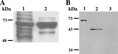
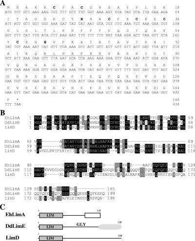
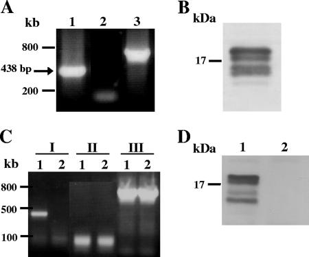
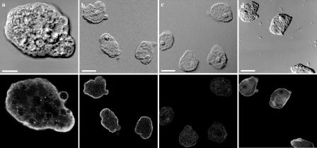
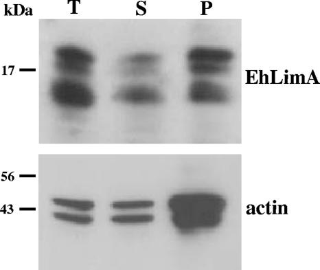
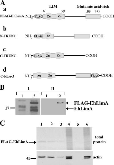
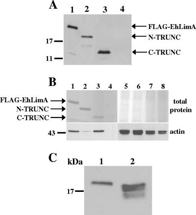
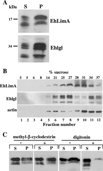
The parasitic protozoan Entamoeba histolytica relies on a very dynamic cytoskeleton in order to invade and survive in host tissues. Identification of cytoskeletal elements is key to understanding these processes. Here we present the characterization of EhLimA, the first LIM protein of E. histolytica. EhLimA consists of a single LIM domain at its N terminus and exhibits the highest degree of homology with DdLimE from Dictyostelium discoideum. Immunofluorescence localization of EhLimA using anti-EhLimA antibodies revealed that EhLimA is highly concentrated at the plasma membrane of cells. Silencing or overexpression of the EhLimA gene did not have a significant effect on the growth or morphology of the parasite. EhLimA associates with the cytoskeleton as demonstrated by the enrichment of the protein in cytoskeleton fractions as well as in pull-down assays that revealed that cytoskeleton association involves interaction with actin. EhLimA binding to actin was shown to be dependent on the N-terminal LIM domain of EhLimA, as removal of even half of the LIM domain resulted in almost complete inhibition of the binding to actin. We also found that a portion of EhLimA floats to the lower-density regions of a sucrose gradient together with portions of the Gal-lectin light subunit and actin. Treatment of cells with the cholesterol-sequestering agent digitonin resulted in increased solubility of EhLimA. These results indicate that in addition to cytoskeletal association, EhLimA may also associate with lipid rafts in the parasite plasma membrane and suggest that EhLimA may be part of the molecular system connecting the actin cytoskeleton to membrane rafts.


Figures

FIG. 1.
FIG. 1.
(A) Analysis of the His-tagged xylanase-EhLimA by gel electrophoresis following expression and purification in E. coli. Lane 1, total lysate of bacterial cells overexpressing the His-tagged xylanase-EhLimA fusion protein; lane 2, purified xylanase-EhLimA fusion protein. (B) A Western blot analysis demonstrating the specificity of the purified anti-EhLimA antibodies. The purified xylanase-EhLimA fusion protein (lane 1) and a purified glutathione S-transferase (GST)-EhLimA fusion protein (lane 2) are recognized by the antibodies, while purified GST (lane 3) is not.
FIG. 2.
FIG. 2.
(A) Nucleotide sequence and deduced amino acid sequence of EhLimA. The conserved cysteine and histidine residues of the N-terminal LIM domain are shown (bold letters). The glutamic acid-rich C terminus of the protein is indicated by brackets. Part of the repeat motif that appears twice in Dictyostelium DdLimE is underlined. (B) Alignment of the EhLimA amino acid sequence with the amino acid sequences of Dictyostelium DdLimE and LimD. Identical residues are represented by black boxes, and similar residues are represented by gray boxes. Sequences were aligned using the ClustalW multiple-sequence alignment program (www.ebi.ac.uk/clustalw/). GenBank accession numbers are as follows: EhLimA, XP_656918; DdLimE, U97699; LimD, AF348467. (C) Schematic representation of EhLimA and Dictyostelium proteins DdLimE and LimD. The single N-terminal LIM domain present in all proteins is indicated. The glutamic acid-rich region of EhLimA is represented by a white box. The glycine-rich region (GLY) and coiled-coil region (shaded oval) of DdLimE are shown.
FIG. 3.
FIG. 3.
mRNA and protein expression of EhLimA. (A) An RT-PCR analysis of total RNA using primers specific for EhLimA results in amplification of a band of the expected size of 438 bp (lane 1). No amplification is attained when the reverse transcription reaction is performed without reverse transcriptase (lane 2). Amplification of actin is shown in lane 3. (B) Detection of EhLimA in a Western blot analysis of total cell lysates using purified anti-EhLimA antibodies reveals four bands around 16 kDa. (C) An RT-PCR analysis of total RNA of G3 transfected trophozoites in which EhLimA is transcriptionally silenced (lane 2) and of nontransfected G3 cells expressing EhLimA (lane 1). Amplification of EhLimA (lanes I) is attained only with the nontransfected G3 cells but not with the EhLimA-silenced cells. No amplification is attained when the reverse transcription reaction is performed without reverse transcriptase (lanes II). Amplification of actin (lanes III) is attained with both the nontransfected G3 cells and with the EhLimA-silenced cells. (D) Western blot analysis of total cell lysates of G3 transfected trophozoites in which EhLimA is transcriptionally silenced. The anti-EhLimA antibodies do not detect any protein in these cells (lane 2), whereas the protein is detected in the nontransfected G3 cells expressing EhLimA (lane 1).
FIG. 4.
FIG. 4.
Immunofluorescence localization of EhLimA in whole parasites. Trophozoites were fixed and stained for immunofluorescence with anti-EhLimA antibodies followed by fluorescein isothiocyanate-conjugated anti-rabbit secondary antibodies and viewed under a confocal microscope. The differential interference contrast images are shown on the top, and the corresponding fluorescence images are shown on the bottom. EhLimA localizes mainly to the plasma membrane of cells (a and b). No staining was detected in the EhLimA-silenced G3 amoeba cells (c). Staining cells with phalloidin-TRITC reveals and enrichment of actin in the cell cortex and in cellular protrusions (d). Bars, 10 μm in panel a and 20 μm in panels b, c, and d.
FIG. 5.
FIG. 5.
EhLimA associates with the cytoskeleton. Total cell lysates (T) prepared with the addition of Triton X-100 were subjected to ultracentrifugation resulting in a pellet fraction (P) containing the Triton X-100-insoluble cytoskeleton and a supernatant fraction (S) containing soluble proteins. A Western blot analysis of these fractions using anti-EhLimA antibodies shows that a significant portion of EhLimA is found in the Triton X-100-insoluble cytoskeleton fraction. Monoclonal antiactin antibodies reveal that the majority of actin is detected in this fraction as well.
FIG. 6.
FIG. 6.
(A) Schematic representation of constructs encoding full-length and truncated FLAG-tagged EhLimA. (a) N-terminus-tagged full-length EhLimA (FLAG-EhLimA); (b) N-terminus-tagged EhLimA lacking half of the N terminus LIM domain (N-TRUNC); (c) N-terminus-tagged EhLimA lacking the glutamic acid-rich C terminus (C-TRUNC); (d) C-terminus-tagged full-length EhLimA (C-FLAG). (B) Protein expression of FLAG-EhLimA is confirmed in a Western blot analysis using anti-EhLimA antibodies (lanes I) and anti-FLAG monoclonal antibodies (lanes II). Lane 1 and 2 contain total cell lysates of wild-type cells and FLAG-EhLimA-overexpressing cells, respectively. (C) Different amounts of total cell lysates of FLAG-EhLimA-overexpressing cells (lanes 1 to 3) and wild-type cells (lane 5) were incubated with anti-FLAG monoclonal antibodies covalently attached to agarose. Immunoprecipitated FLAG-EhLimA was analyzed by gel electrophoresis followed by total protein staining of the gel (top gel). Coimmunoprecipitation of actin was analyzed in a Western blot of immunoprecipitated protein products using antiactin antibodies (bottom gel). Lane 1, 250 μg total lysate; lane 2, 100 μg total lysate; lane 3, 50 μg total lysate; lane 5, 500 μg total lysate. Protein amounts are confirmed in lanes 4 and 6 for FLAG-EhLimA-overexpressing cells and wild-type cells, respectively, each containing 10 μg of total lysate.
FIG. 7.
FIG. 7.
(A) Expression of N-terminus- and C-terminus-truncated FLAG-tagged EhLimA. Protein expression of N-terminus-truncated FLAG-tagged EhLimA (N-TRUNC, lane 2) and C-terminus-truncated FLAG-tagged EhLimA (C-TRUNC, lane 3) is confirmed in a Western blot analysis with anti-FLAG antibodies. In lane 1, expression of the full-length FLAG-EhLimA fusion protein is shown. Lane 4 contains a total cell lysate of wild-type cells. (B) Immunoprecipitation and actin binding of N-terminus- and C-terminus-truncated FLAG-tagged EhLimA. Total cell lysates of cells overexpressing FLAG- EhLimA (lane 1), N-TRUNC (lane 2), or C-TRUNC (lane 3) and wild-type cells (lane 4) were incubated with anti-FLAG monoclonal antibodies covalently attached to agarose. Immunoprecipitated proteins were analyzed by gel electrophoresis, followed by total protein staining of the gel (top gel). A Western blot analysis of immunoprecipitated protein products using antiactin antibodies reveals that N-TRUNC has almost completely lost the ability to bind actin (bottom gel). Protein amounts are confirmed in lanes 5 to 8 for lanes 1 to 4, respectively, each containing 10 μg of total lysate. (C) N-terminal cleavage of EhLimA. Anti-FLAG antibodies detect four bands in a Western blot of total cell lysates of cells overexpressing FLAG-tagged full-length EhLimA in which the FLAG epitope is located at the C terminus of the protein (C-FLAG, lane 2) as opposed to only one band that is detected in total cell lysates of the N-terminus-tagged FLAG-EhLimA-overexpressing cells (lane 1).
FIG. 8.
FIG. 8.
A portion of EhLimA associates with lipid rafts. (A) Total cell lysates prepared with the addition of cold Triton X-100 were subjected to centrifugation resulting in soluble (S) and insoluble (P) fractions. A Western blot analysis using anti-EhLimA antibodies shows that a significant portion of EhLimA is found in the Triton X-100-insoluble fraction. A similar protein distribution of the amoebic Gal-lectin light subunit (Ehlgl) was detected using polyclonal anti-Ehlgl antibodies. (B) Total cell lysates prepared with cold Triton X-100 were subjected to sucrose gradient flotation. Twelve fractions were collected from the top of the gradient and proteins were precipitated with trichloroacetic acid. Following SDS-PAGE and immunoblotting, the distribution of EhLimA, Ehlgl, and actin in the fractions was analyzed. A portion of EhLimA as well as of Ehlgl and actin was detected in the lower-density regions of the gradient. The presence of EhLimA in the lower-density regions was observed in three independent experiments. The percentage of sucrose in each fraction is indicated. (C) Depletion of cholesterol. Cells were incubated for 30 min with either digitonin or methyl-β-cyclodextrin on ice followed by lysis in Triton X-100 and centrifugation. Western blotting reveals that the distribution of EhLimA between soluble (S) and insoluble (P) fractions was greatly affected by digitonin but not by methyl-β-cyclodextrin.

Similar articles

Cited by

References

    1. Abrami, L., and F. G. van Der Goot. 1999. Plasma membrane microdomains act as concentration platforms to facilitate intoxication by aerolysin. J. Cell Biol. 147:175-184. - PMC - PubMed
    1. Algrain, M., O. Turunen, A. Vaheri, D. Louvard, and M. Arpin. 1993. Ezrin contains cytoskeleton and membrane binding domains accounting for its proposed role as a membrane-cytoskeletal linker. J. Cell Biol. 120:129-139. - PMC - PubMed
    1. Ankri, S., F. Padilla-Vaca, T. Stolarsky, L. Koole, U. Katz, and D. Mirelman. 1999. Antisense inhibition of expression of the light subunit (35 kDa) of the Gal/GalNac lectin complex inhibits Entamoeba histolytica virulence. Mol. Microbiol. 33:327-337. - PubMed
    1. Bach, I. 2000. The LIM domain: regulation by association. Mech. Dev. 91:5-17. - PubMed
    1. Baltz, R., C. Domon, D. T. Pillay, and A. Steinmetz. 1992. Characterization of a pollen-specific cDNA from sunflower encoding a zinc finger protein. Plant J. 2:713-721. - PubMed

Publication types