. 2007 Aug 7;46(31):9133-42.
doi: 10.1021/bi700944j. Epub 2007 Jul 13.

Direct evidence for specific interactions of the fibrinogen alphaC-domains with the central E region and with each other

Affiliations

Direct evidence for specific interactions of the fibrinogen alphaC-domains with the central E region and with each other

Rustem I Litvinov et al. Biochemistry. .

Abstract

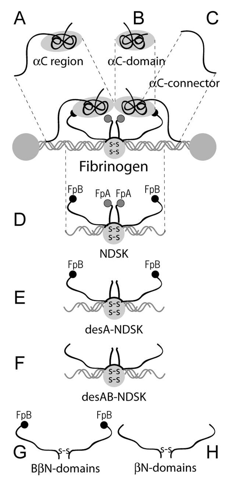
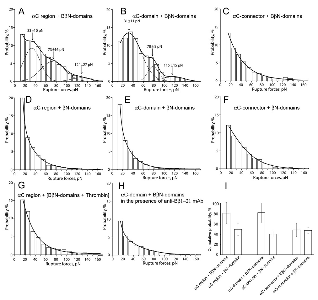
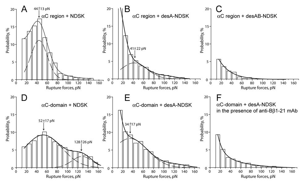
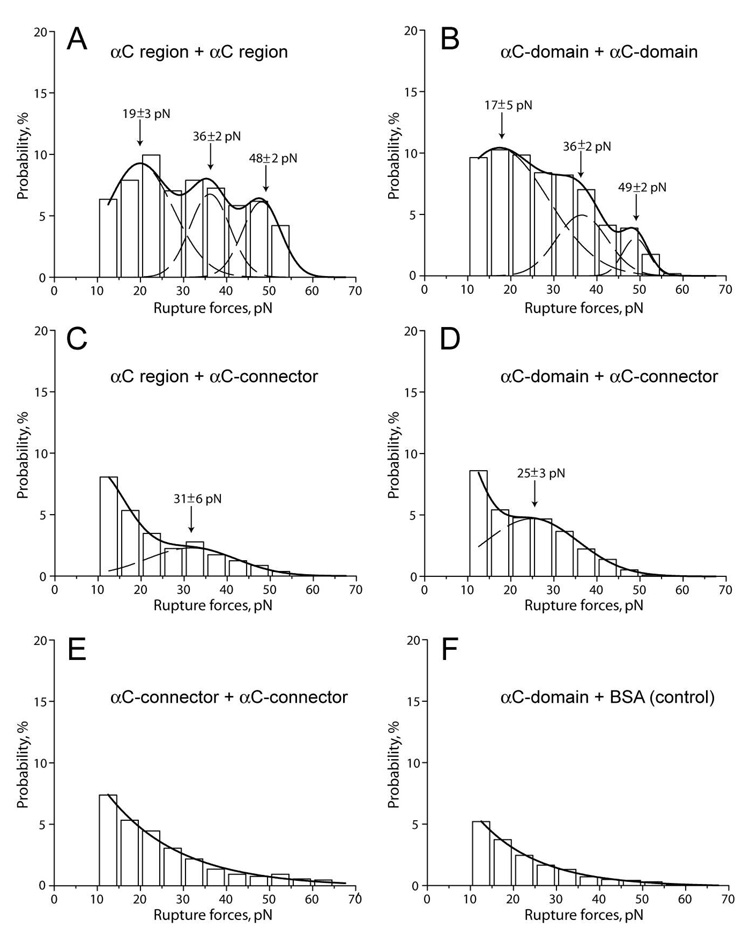
The carboxyl-terminal regions of the fibrinogen Aalpha chains (alphaC regions) form compact alphaC-domains tethered to the bulk of the molecule with flexible alphaC-connectors. It was hypothesized that in fibrinogen two alphaC-domains interact intramolecularly with each other and with the central E region preferentially through its N-termini of Bbeta chains and that removal of fibrinopeptides A and B upon fibrin assembly results in dissociation of the alphaC regions and their switch to intermolecular interactions. To test this hypothesis, we studied the interactions of the recombinant alphaC region (Aalpha221-610 fragment) and its subfragments, alphaC-connector (Aalpha221-391) and alphaC-domain (Aalpha392-610), between each other and with the recombinant (Bbeta1-66)2 and (beta15-66)2 fragments and NDSK corresponding to the fibrin(ogen) central E region, using laser tweezers-based force spectroscopy. The alphaC-domain, but not the alphaC-connector, bound to NDSK, which contains fibrinopeptides A and B, and less frequently to desA-NDSK and (Bbeta1-66)2 containing only fibrinopeptides B; it was poorly reactive with desAB-NDSK and (beta15-66)2 both lacking fibrinopeptide B. The interactions of the alphaC-domains with each other and with the alphaC-connector were also observed, although they were weaker and heterogeneous in strength. These results provide the first direct evidence for the interaction between the alphaC-domains and the central E region through fibrinopeptide B, in agreement with the hypothesis given above, and indicate that fibrinopeptide A is also involved. They also confirm the hypothesized homomeric interactions between the alphaC-domains and display their interaction with the alphaC-connectors, which may contribute to covalent cross-linking of alpha polymers in fibrin.


Figures

Figure 1
Figure 1. Cartoon of fibrinogen and fibrin(ogen) fragments used in this study
A, B, and C show the αC region fragment corresponding to the C-terminal portion of the fibrinogen Aα chain (residues Aα221-610) and its sub-fragments, the αC-domain (residues Aα392-610) and αC-connector (residues Aα221-391), respectively. D, E, and F show NDSK [“N-Terminal DiSulphide Knot”(3)], a fragment from the central part of fibrinogen containing both FpA and FpB, desA-NDSK with cleaved FpA but remaining FpB, and desAB-NDSK with both FpA and FpB cleaved, respectively. G shows the recombinant fibrinogen (Bβ1-66)2 fragment consisting of two BβN-domains formed by the N-terminal portions of the fibrinogen Bβ chain. H shows the recombinant fibrin fragment (β15-66)2 including two βN-domains devoid of fibrinopeptides B. The grey and black circles on the ends represent fibrinopeptides A (FpA) and B (FpB), respectively. The gray circle in the center with double designations “S-S” inside represents a cluster of disulphide bonds.
Figure 2
Figure 2. The panel of rupture force histograms demonstrating interactions of the recombinant fragment corresponding to the αC region and its sub-fragments, αC-connector and αC-domain, with the recombinant (Bβ1-66)2 and (β15-66)2 fragments corresponding to the fibrinogen BβN- and fibrin βN-domains, respectively
A, B, and C – interactions of the BβN-domains with the αC region, αC-domain, and αC-connector, respectively; D, E, and F - interactions of the βN-domains with the αC region, αC-domain, and αC-connector, respectively; G - interactions of the αC region with the BβN-domains treated with thrombin and thus converted to the βN-domains right on the surface; H - interactions of the αC-domain with the BβN-domains in the presence of 200 µg/ml anti-Bβ1-21 mAb; I – paired bars representing cumulative probabilities of forces >10 pN derived from A and D, B and E, and C and F. The dashed lines show the fitting with Gaussian curves to determine the position of each peak that corresponds to the most probable rupture force.
Figure 3
Figure 3. The panel of rupture force histograms demonstrating interactions of the recombinant αC region and αC-domain with various NDSK fragments corresponding to the central E region of fibrin(ogen)
A, B, and C - interactions of the αC region with NDSK, desA-NDSK, and desAB-NDSK, respectively; D and E - interactions of the αC-domain with NDSK and desA-NDSK, respectively; F – the same as in E, but in the presence of 200 µg/ml anti-Bβ1-21 mAb. The dashed lines show the fitting with Gaussian curves to determine the position of each peak that corresponds to the most probable rupture force.
Figure 4
Figure 4. The panel of rupture force histograms demonstrating interactions of the αC region and its sub-fragments, αC-domain and αC-connector
A - interactions of the pedestal-bound αC region with the αC region coupled to a bead; B - the pedestal-bound αC-domain with the αC-domain coupled to a bead; C, D, and E – the pedestal-bound αC region, αC-domain, and αC-connector with the αC-connector coupled to a bead, respectively; F - the pedestal-bound αC-domain with the BSA-coated bead (negative control). The dashed lines show the fitting with Gaussian curves to determine the position of each peak that corresponds to the most probable rupture force.

References

    1. Weisel JW. Fibrinogen and fibrin. Adv. Protein Chem. 2005;70:247–299. - PubMed
    1. Mosesson MW. Fibrinogen and fibrin structure and functions. J Thromb Haemost. 2005;3:1894–1904. - PubMed
    1. Blomback B, Blomback M, Henschen A, Hessel B, Iwanaga S, Woods KR. N-terminal disulphide knot of human fibrinogen. Nature. 1968;218:130–134. - PubMed
    1. Brown JH, Volkmann N, Jun G, Henschen-Edman AH, Cohen C. The crystal structure of modified bovine fibrinogen. Proc Natl Acad Sci U S A. 2000;97:85–90. - PMC - PubMed
    1. Yang Z, Kollman JM, Pandi L, Doolittle RF. Crystal structure of native chicken fibrinogen at 2.7 A resolution. Biochemistry. 2001;40:12515–12523. - PubMed

Publication types

MeSH terms