

Identification and metabolic transformations of carotenoids in ocular tissues of the Japanese quail Coturnix japonica
- PMID: 17630780
- PMCID: PMC2531157
- DOI: 10.1021/bi700558f
Identification and metabolic transformations of carotenoids in ocular tissues of the Japanese quail Coturnix japonica
Abstract
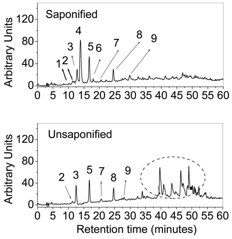
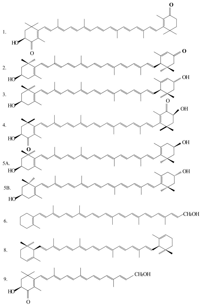
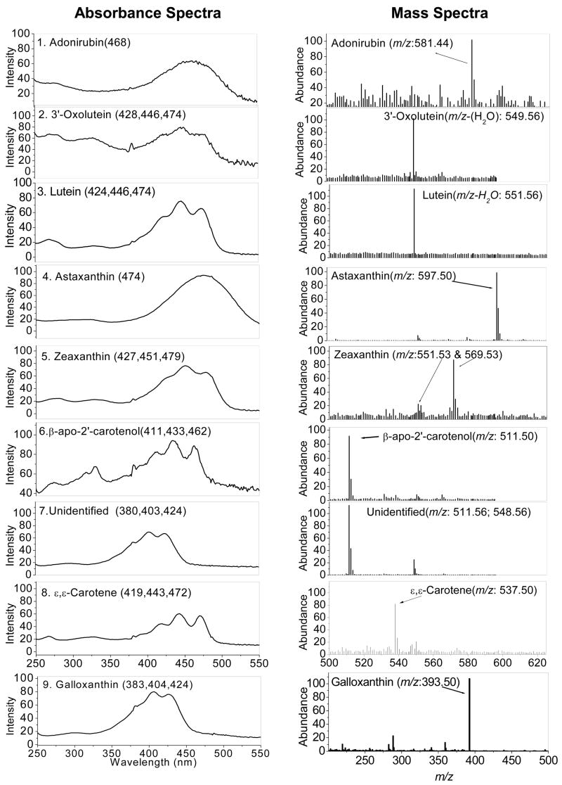
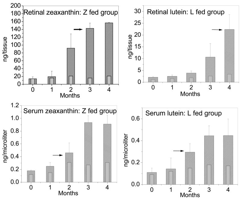
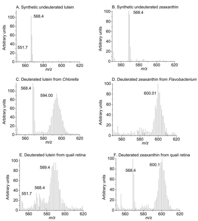
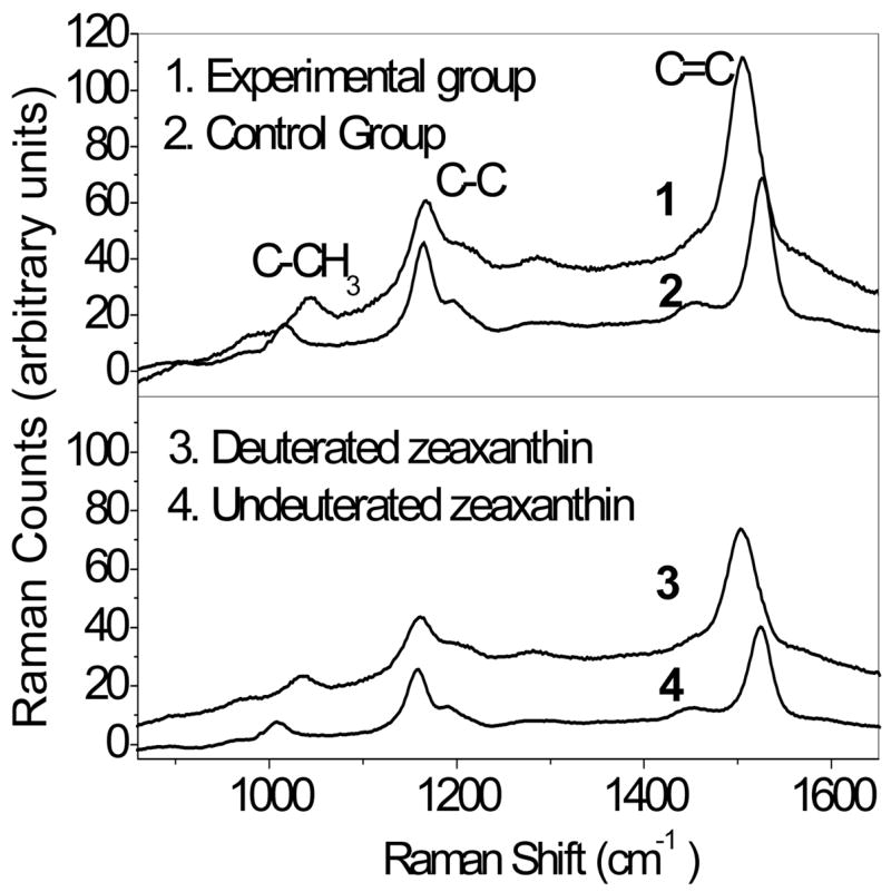
As in humans and monkeys, lutein [(3R,3'R,6'R)-beta,epsilon-carotene-3,3'-diol] and zeaxanthin [a mixture of (3R,3'R)-beta,beta-carotene-3,3'diol and (3R,3'S-meso)-beta,beta-carotene-3,3'-diol] are found in substantial amounts in the retina of the Japanese quail Coturnix japonica. This makes the quail retina an excellent nonprimate small animal model for studying the metabolic transformations of these important macular carotenoids that are thought to play an integral role in protection against light-induced oxidative damage such as that found in age-related macular degeneration (AMD). In this study, we first identified the array of carotenoids present in the quail retina using C30 HPLC coupled with in-line mass spectral and photodiode array detectors. In addition to dietary lutein (2.1%) and zeaxanthin (11.8%), we identified adonirubin (5.4%), 3'-oxolutein (3.8%), meso-zeaxanthin (3.0%), astaxanthin (28.2%), galloxanthin (12.2%), epsilon,epsilon-carotene (18.5%), and beta-apo-2'-carotenol (9.5%) as major ocular carotenoids. We next used deuterium-labeled lutein and zeaxanthin as dietary supplements to study the pharmacokinetics and metabolic transformations of these two ocular pigments in serum and ocular tissues. We then detected and quantitated labeled carotenoids in ocular tissue using both HPLC-coupled mass spectrometry and noninvasive resonance Raman spectroscopy. Results indicated that dietary zeaxanthin is the precursor of 3'-oxolutein, beta-apo-2'-carotenol, adonirubin, astaxanthin, galloxanthin, and epsilon,epsilon-carotene, whereas dietary lutein is the precursor for meso-zeaxanthin. Studies also revealed that the pharmacokinetic patterns of uptake, carotenoid absorption, and transport from serum into ocular tissues were similar to results observed in most human clinical studies.
Figures
References
-
- Mares-Perlman JA, Millen AE, Ficek TL, Hankinson SE. The body of evidence to support a protective role for lutein and zeaxanthin in delaying chronic disease. Overview, J Nutr. 2002;132:518S–524S. - PubMed
-
- Gale CR, Hall NF, Phillips DI, Martyn CN. Lutein and zeaxanthin status and risk of age-related macular degeneration. Invest Ophthalmol Vis Sci. 2003;44:2461–2465. - PubMed
-
- Krinsky NI, Johnson EJ. Carotenoid actions and their relation to health and disease. Mol Aspects Med. 2005;26:459–516. - PubMed
-
- Krinsky NI, Landrum JT, Bone RA. Biologic mechanisms of the protective role of lutein and zeaxanthin in the eye. Annu Rev Nutr. 2003;23:171–201. - PubMed
-
- Seddon JMAU, Sperduto RD, Hiller R, Blair N, Burton TC, Farber MD, Gragoudas ES, Haller J, Miller DT, et al. Dietary carotenoids, vitamins A, C, and E, and advanced age-related macular degeneration. Eye Disease Case-Control Study Group. JAMA. 1994;272:1413–1420. - PubMed
Publication types
MeSH terms
Substances
Grants and funding
LinkOut - more resources
Full Text Sources
Medical
Research Materials
