. 2007 Jul;3(7):e85.
doi: 10.1371/journal.ppat.0030085.

HIV-1 Vpr-mediated G2 arrest involves the DDB1-CUL4AVPRBP E3 ubiquitin ligase

Affiliations

HIV-1 Vpr-mediated G2 arrest involves the DDB1-CUL4AVPRBP E3 ubiquitin ligase

Jean-Philippe Belzile et al. PLoS Pathog. 2007 Jul.

Abstract

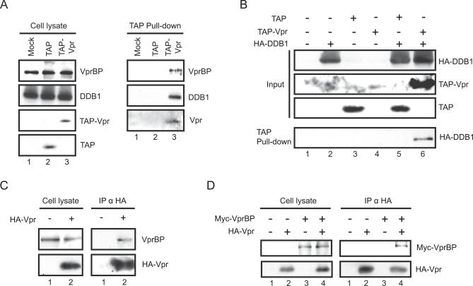
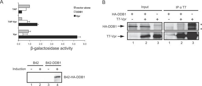
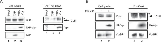
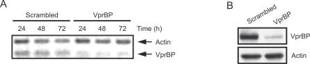
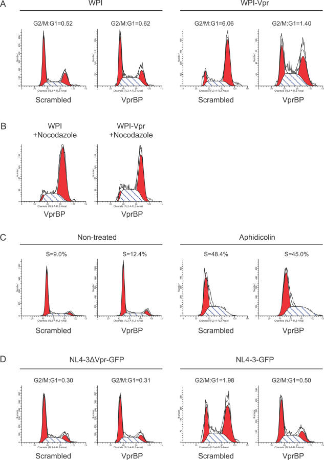
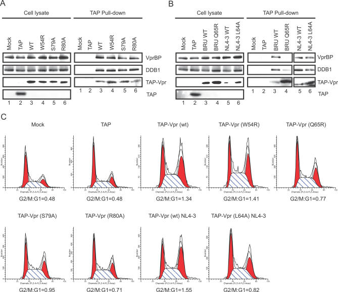
Human immunodeficiency virus type 1 (HIV-1) viral protein R (Vpr) has been shown to cause G2 cell cycle arrest in human cells by inducing ATR-mediated inactivation of p34cdc2, but factors directly engaged in this process remain unknown. We used tandem affinity purification to isolate native Vpr complexes. We found that damaged DNA binding protein 1 (DDB1), viral protein R binding protein (VPRBP), and cullin 4A (CUL4A)--components of a CUL4A E3 ubiquitin ligase complex, DDB1-CUL4A(VPRBP)--were able to associate with Vpr. Depletion of VPRBP by small interfering RNA impaired Vpr-mediated induction of G2 arrest. Importantly, VPRBP knockdown alone did not affect normal cell cycle progression or activation of ATR checkpoints, suggesting that the involvement of VPRBP in G2 arrest was specific to Vpr. Moreover, leucine/isoleucine-rich domain Vpr mutants impaired in their ability to interact with VPRBP and DDB1 also produced strongly attenuated G2 arrest. In contrast, G2 arrest-defective C-terminal Vpr mutants were found to maintain their ability to associate with these proteins, suggesting that the interaction of Vpr with the DDB1-VPRBP complex is necessary but not sufficient to block cell cycle progression. Overall, these results point toward a model in which Vpr could act as a connector between the DDB1-CUL4A(VPRBP) E3 ubiquitin ligase complex and an unknown cellular factor whose proteolysis or modulation of activity through ubiquitination would activate ATR-mediated checkpoint signaling and induce G2 arrest.


Conflict of interest statement

Competing interests. The authors have declared that no competing interests exist.

Figures

Figure 1
Figure 1. Immunoprecipitation of DDB1/Vpr and VPRBP/Vpr Complexes
(A) HEK293T cells were mock transfected (lanes 1) or transfected with either TAP (lanes 2) or TAP-Vpr–expressing plasmids (lanes 3). Two days later, immunoprecipitations of TAP tag were performed on cell lysates using IgG-coupled beads and purified complexes were eluted by cleavage with TEV protease. The levels of endogenous VPRBP and DDB1 were monitored in crude lysates and pulled-down fractions by Western blot using specific antibodies. TAP, TAP-Vpr, and cleaved Vpr were detected using a polyclonal rabbit antibody directed against a Vpr N-terminal peptide. (B) HEK293T cells were mock transfected (lanes 1 and 2) or transfected with either TAP (lanes 3 and 5) or TAP-Vpr–expressing plasmids (lanes 4 and 6). Cells were transcomplemented with the empty vector (lanes 1, 3, and 4) or HA-DDB1–encoding plasmid (lanes 2, 5, and 6). (C) HEK293T cells were mock transfected (lanes 1) or transfected with HA-Vpr–expressing plasmid (lanes 2). Immunoprecipitations using anti-HA antibodies were performed on cell extracts using protein A–sepharose beads. The levels of HA-Vpr and endogenous VPRBP were monitored in cell extracts as well as immunoprecipitated fractions by Western blot using specific antibodies. (D) HEK293T cells were mock transfected (lanes 1 and 3) or transfected with a HA-Vpr–expressing plasmid (lanes 2 and 4). Cells were transcomplemented with the empty vector (lanes 1 and 2) or Myc-VPRBP–encoding plasmid (lanes 3 and 4). Anti-HA immunoprecipitations were performed as described above.
Figure 2
Figure 2. Absence of Direct Vpr Binding to DDB1 in Yeast
(A) The EGY48 reporter strain containing LexA-TAP, LexA-Vpr, or LexA-TAP-Vpr (“bait”) was transformed with B42, B42-DDB1, or B42-Vpr–expressing plasmid (“prey”). The binding affinity between the different proteins was assessed by assaying β-galactosidase activity using the o-nitrophenyl-β-D-galactopyranoside method. Histograms represent averaged data from 2–4 different clones and are representative of two independent assays. Western blot analysis of induced and non-induced B42-HA-DDB1 expression in the B42 and B42-DDB1-transformed EGY48/LexA-Vpr reporter strain is shown below. (B) In vitro–translated T7-Vpr was immunoprecipitated with an anti-T7 antibody in the presence or absence of in vitro–translated HA-DDB1. Amounts of protein initially added to the assay (input) are shown in the left panel. * represents non-specific proteins immunoprecipitated by the anti-T7 antibody.
Figure 3
Figure 3. Vpr Associates with the Ubiquitin Ligase Scaffold Protein CUL4A
(A) Five million HEK293T cells were transfected with 40 μg of empty (lanes 1), TAP-expressing (lanes 2), or TAP-Vpr–expressing plasmids (lanes 3). Forty-eight hours after transfection, TAP pull-downs were performed on cell lysates using IgG-coupled beads, and purified complexes were eluted by cleavage with TEV protease. The levels of endogenous CUL4A were monitored in crude lysates and pulled-down fractions by Western blot using a polyclonal goat anti-CUL4A antibody. TAP, TAP-Vpr, and cleaved Vpr were detected using a polyclonal rabbit antibody directed against a Vpr N-terminal peptide. (B) Ten million HEK293T cells were transfected with 80 μg of empty plasmid (lanes 1) or with HA-Vpr–expressing plasmid (lanes 2). Immunoprecipitation of endogenous CUL4A was performed using a goat polyclonal anti-CUL4A antibody and protein A–sepharose beads. The levels of endogenous CUL4A, VPRBP, and over-expressed HA-Vpr were monitored in crude lysates and immunoprecipitated fractions by Western blot using, respectively, a polyclonal goat anti-CUL4A antibody, a polyclonal rabbit anti-VPRBP antibody, and a monoclonal mouse anti-HA antibody. * represents a non-specific protein detected by the anti-CUL4A antibody. The anti-CUL4A antibody generally recognized a doublet of CUL4A when the gel resolution was sufficiently high. In the TAP pull-down fractions, only the upper band of CUL4A was detected.
Figure 4
Figure 4. Depletion of VPRBP Using siRNA
HEK293 cells were transfected with 300 pmol of VPRBP-targeting siRNA or control scrambled siRNA using Oligofectamine. (A) At 24, 48, and 72 h post-transfection, RNA was extracted and analyzed by RT-PCR to determine the extent of VPRBP downregulation at the mRNA level. PCR products were analyzed in the exponential phase of amplification. Actin levels were used as a control for RNA quality and reverse transcription efficiency. (B) Forty-eight hours after siRNA transfection, cells lysates were harvested and analyzed by Western blot using a polyclonal rabbit anti-VPRBP antibody to demonstrate the downregulation of VPRBP at the protein level. Actin levels were used as a control for protein loading.
Figure 5
Figure 5. Effect of VPRBP Depletion on Vpr-Induced G2 Arrest
(A) HEK293 cells were transfected with 300 pmol of VPRBP-targeting siRNA or control scrambled siRNA using Oligofectamine, followed by the same transfection 24 h later. Twenty-four hours after the second siRNA transfection, cells were transduced at a multiplicity of infection of 1 with lentiviral vectors expressing Vpr (WPI-Vpr) or the empty vector (WPI). Cell cycle profiles were analyzed 24 h after transduction by flow cytometry using propidium iodide staining. (B and C) To determine if cell growth (B) or checkpoint activation (C) was affected by VPRBP knockdown, HEK293 cells were transfected once with siRNA, as described above, and treated respectively with 1 μg/ml nocodazole and 0.5 μM aphidicolin 24 h later. Cell cycle profiles were analyzed 24 h after drug treatment. (D) To determine if VPRBP knockdown could also abrogate the induction of G2 arrest in the context of viral infection, siRNA-transfected cells were infected with NL4.3-GFP and NL4–3ΔVpr-GFP at a concentration of 100 cpm/cell and cell cycle profiles were analyzed 48 h later. Percentages of G1 and G2/M cell populations were determined using the ModFit software. These results are representative of the data obtained in at least two independent experiments.
Figure 6
Figure 6. DDB1 and VPRBP Binding Affinities of TAP-Tagged Vpr Mutants
(A) HEK293T cells were transfected with TAP-Vpr plasmids encoding for wild-type Vpr (lanes 3) or Vpr mutants W54R (lanes 4), S79A (lanes 5), and R80A (lanes 6). As control, cells were mock transfected (lanes 1) or transfected with a TAP-expressing plasmid (lanes 2). Following TAP pull-down using IgG-coupled beads, the levels of endogenous VPRBP and DDB1 were monitored in crude lysates and pulled-down fractions by Western blot using specific antibodies. TAP, TAP-Vpr, and cleaved Vpr were detected using a polyclonal rabbit antibody directed against a Vpr N-terminal peptide. (B) HEK293T cells were transfected with TAP-Vpr plasmids encoding for wild-type Bru Vpr (lanes 3) and wild-type NL4–3 Vpr (lanes 5), or Vpr mutants Bru Q65R (lanes 4) and NL4–3 L64A (lanes 6). As control, cells were mock transfected (lanes 1) or transfected with a TAP-expressing plasmid (lanes 2). TAP pull-downs and immunodetection of VPRBP, DDB1, TAP, and Vpr were performed as described for (A). (C) HEK293T cells were co-transfected with 1 μg of GFP-expressing plasmid and 15 μg of TAP-Vpr plasmids expressing wild-type or mutant proteins. Cell cycle analysis was performed using propidium iodide staining on the GFP+ cell population as described in Materials and Methods. Percentages of G1 and G2/M cell populations were determined using the ModFit software.

References

    1. Le Rouzic E, Benichou S. The Vpr protein from HIV-1: Distinct roles along the viral life cycle. Retrovirology. 2005;2:11. - PMC - PubMed
    1. Andersen JL, Planelles V. The role of Vpr in HIV-1 pathogenesis. Curr HIV Res. 2005;3:43–51. - PubMed
    1. Jowett JB, Planelles V, Poon B, Shah NP, Chen ML, et al. The human immunodeficiency virus type 1 vpr gene arrests infected T cells in the G2 + M phase of the cell cycle. J Virol. 1995;69:6304–6313. - PMC - PubMed
    1. He J, Choe S, Walker R, Di Marzio P, Morgan DO, et al. Human immunodeficiency virus type 1 viral protein R (Vpr) arrests cells in the G2 phase of the cell cycle by inhibiting p34cdc2 activity. J Virol. 1995;69:6705–6711. - PMC - PubMed
    1. Rogel ME, Wu LI, Emerman M. The human immunodeficiency virus type 1 vpr gene prevents cell proliferation during chronic infection. J Virol. 1995;69:882–888. - PMC - PubMed

Publication types

MeSH terms

Substances