

Review
doi: 10.1016/j.dnarep.2007.05.009.
Epub 2007 Jul 12.
The X family portrait: structural insights into biological functions of X family polymerases
Affiliations
- PMID: 17631059
- PMCID: PMC2128704
- DOI: 10.1016/j.dnarep.2007.05.009
Item in Clipboard
Review
The X family portrait: structural insights into biological functions of X family polymerases
DNA Repair (Amst).
.
Abstract
The mammalian family X DNA polymerases (DNA polymerases beta, lambda, mu, and TdT) contribute to base excision repair and double-strand break repair by virtue of their ability to fill short gaps in DNA. Structural information now exists for all four of these enzymes, making this the first mammalian polymerase family whose structural portrait is complete. Here we consider how distinctive structural features of these enzymes contribute to their biological functions in vivo.
Figures
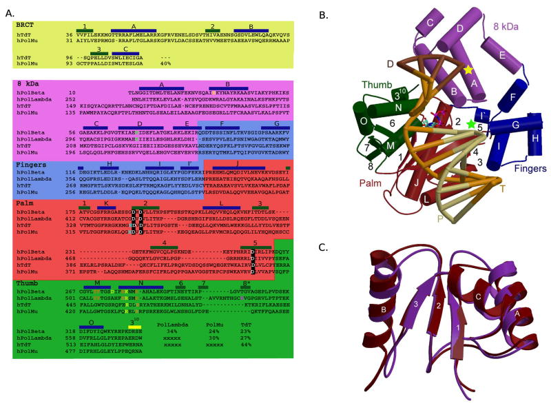
Mammalian Family X polymerases. (A) Structure-based primary sequence alignment. Catalytic aspartates, boxed in black; residues involved in minor groove interactions, orange; residues involved in stacking interactions with nucleotides, yellow; dRP lyase nucleophile, green; active site histidines (Pol μ H329, TdT H342), cyan; residue putatively stabilizing frameshift intermediate, magenta. Secondary structural elements are blue (α-helices), green (β-strands), and yellow (310 helix) boxes. (B) Domain organization. α-helices are depicted as cylinders, and labeled alphabetically. β-strands are shown as directional arrows, labeled numerically. The bound DNA duplex is shown in the binding cleft: template (T), orange; upstream primer (P), khaki; and downstream primer (D), brown. The position of the 3′-OH and downstream 5′-phosphate are marked with green and yellow stars, respectively. The incoming nucleotide is cyan. The ternary complex of Pol β (PDB code 2FMS) was used for this illustration. (C) NMR solution structure of the Pol μ (maroon) and TdT (purple) BRCT domains. α-helices are labeled alphabetically and β-strands numerically. Numbering only applies within the BRCT domains.
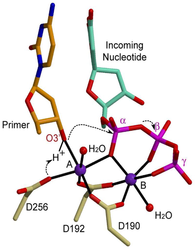
Catalytic mechanism for nucleotidyl transfer. The primer terminal residue (orange) is drawn in stick, with the O3′ in red. The incoming nucleotide is cyan, with its phosphates in magenta. Protein residues from the ternary structure of Pol β (PDB code 2FMS) are khaki. Magnesium ions are purple and water molecules completing coordination of the metal ions are small red spheres.
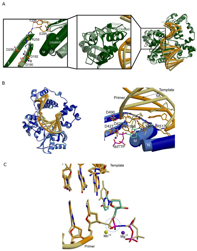
Comparison of binary and ternary complexes for Pol β and Pol λ. (A) Superposition of binary (PDB code 1 BPX; protein, light green; DNA, khaki) and ternary (PDB code 2FMS; protein, dark green; DNA, orange) crystal structures of Pol β (right). Left: Amino acid side chain motions within the active site. Middle: Variations in thumb subdomain position between the ‘open’ (light green) and ‘closed’ (dark green) forms of the enzyme. (B) Superposition of binary (PDB code 1XSL; protein, light blue; DNA, khaki) and ternary (PDB code 1XSN; protein, dark blue; DNA, orange) crystal structures of Pol λ (left). Right: Highlights of amino acid side chain motions in the active site of Pol λ. Binary complex: protein, light blue; DNA khaki. Ternary complex: protein, dark blue; DNA, orange. The incoming nucleotide is drawn in stick (cyan). The magnesium ion in metal site B shown as a purple sphere. (C) Superposition of the pre-catalytic (PDB code 2PFO, orange) and the post-catalytic (PDB code 1XSP, khaki) ternary complexes of Pol λ. The dUMPNPP from the pre-catalytic complex is cyan. The manganese ion in metal site A is shown as a yellow sphere, and the magnesium ion in metal site B is shown as a purple sphere. The α-phosphate undergoing stereochemical inversion is circled in dashed light blue.
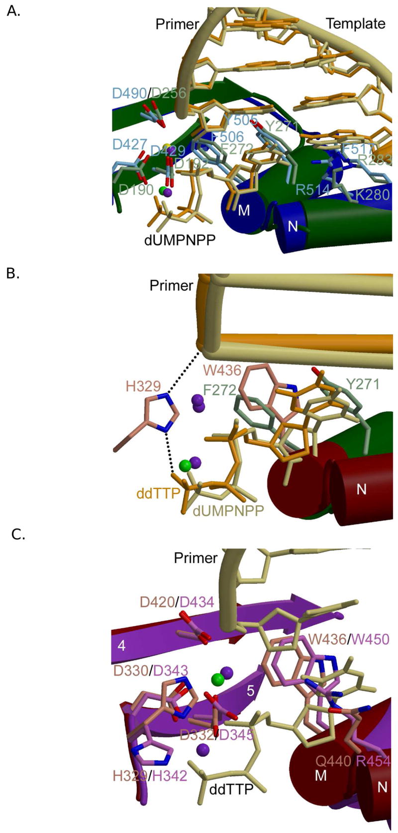
Family X polymerase active site organization. (A) Superposition of ternary complexes of Pol β (PDB code 2FMS; protein, green; DNA, khaki) and Pol λ (PDB code 2PFO; protein, blue; DNA, orange). Protein side chains from Pol β are drawn in light green, while those for Pol λ are light blue. (B) Superposition of the ternary complexes of Pol β (PDB code 2FMS; protein, green; DNA, khaki) and Pol μ (PDB code 2IHM; protein, maroon; DNA, orange). Protein side chains from Pol β are drawn in light green, while those for Pol μ are pink. Putative hydrogen bonding interactions between Pol μ H329 and the primer terminal phosphate or the γ-phosphate of the incoming nucleotide are shown as black dashed lines. Magnesium and sodium ions are shown as purple and green spheres, respectively. (C) Superposition of ternary complexes of Pol μ (PDB code 2IHM; protein, maroon; DNA, khaki) and TdT apoprotein (PDB code 1JMS; protein, purple). Protein side chains from Pol μ are drawn in pink, while those for TdT are light purple. The incoming nucleotide from the structure of Pol μ is drawn in khaki. Magnesium and sodium ions are shown as purple and green spheres, respectively.
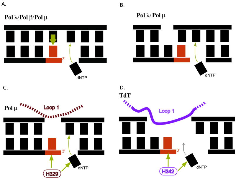
Schematic representation of substrate preferences of Family X polymerases. The primer terminal residue is red and the incoming nucleotide is black. (A) Pol β, Pol λ, and Pol μ can fill small single-strand gaps in a template-dependent manner. (B) Pol λ and Pol μ can fill double-strand gaps with at least one complementary base pair proximal to the gap to be filled. (C) Pol μ, aided by Loop I and H329, can fill a gap from a primer terminal residue with no complementary template strand nucleotide opposite that position. (D) TdT, with the aid of Loop I and H342, utilizes DNA substrates with a 3′-primer terminal overhang, and carries out polymerization in a template-independent manner. Pol μ is also capable of template-independent synthesis in this fashion, but to a lesser extent.
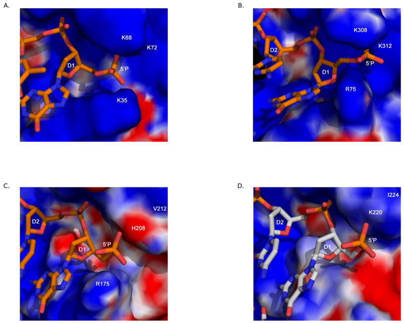
Comparison of the 8 kDa domain 5′-phosphate binding pocket. Electrostatic surface potential plots were calculated using the Adaptive Poisson-Boltzmann Solver tool in PyMOL [89]. The downstream primer is drawn in orange. All DNA and protein residues are labeled in white. The 5′-phosphate binding pocket in the 8 kDa domain of: (A) Pol β (PDB code 2FMS). (B) Pol λ (PDB code 1XSN). (C) Pol μ (PDB code 2IHM). (D) TdT (PDB code 1JMS). The downstream primer from PDB code 2IHM was modeled into the 5′-phosphate binding pocket of TdT, and is drawn in light gray.
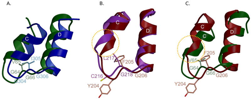
Comparing the 8 kDa domain Helix-hairpin-Helix motifs. (A) HhH motifs from the 8 kDa domains of Pol β (green) and Pol λ (blue) are shown in a ribbon diagram. Side chains in the hairpins of Pol β and Pol λ are light green and light blue, respectively. (B) HhH motifs from the 8 kDa domains of Pol μ (maroon) and TdT (purple). Side chains in the hairpins of Pol μ and TdT are pink and light purple, respectively. (C) HhH motifs from the 8 kDa domains of Pol μ (maroon) and Pol β (green). Side chains from residues in the hairpins of Pol μ and Pol β are pink and light green, respectively. Distortions in α-helix C are highlighted by a dashed circle (orange).
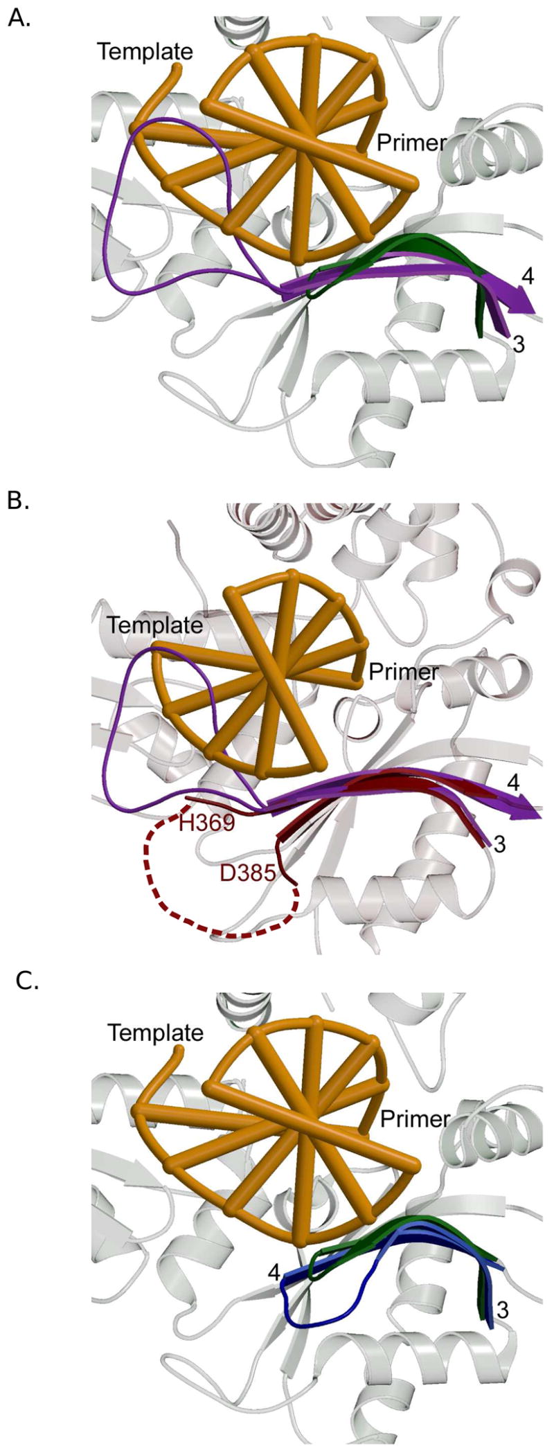
Comparing Loop I in Family X polymerases. Secondary structural elements of Pol β are shown in the background in light gray. The DNA is orange. β-strands 3 and 4 are colored ribbons and labeled in black. (A) Superposition showing the Loop I region of Pol β (PDB code 2FMS, green) and TdT (PDB code 1JMS, purple). (B) Superposition showing the Loop I region of TdT (PDB code 1JMS, purple) and Pol μ (PDB code 2IHM, maroon). Loop I is disordered in Pol μ. The dashed maroon line shows a hypothetical path of the loop in relation to the DNA binding cleft. (C) Superposition of ternary complexes showing the Loop I region of Pol β (PDB code 2FMS, green) and Pol λ (PDB code 1XSN, blue).
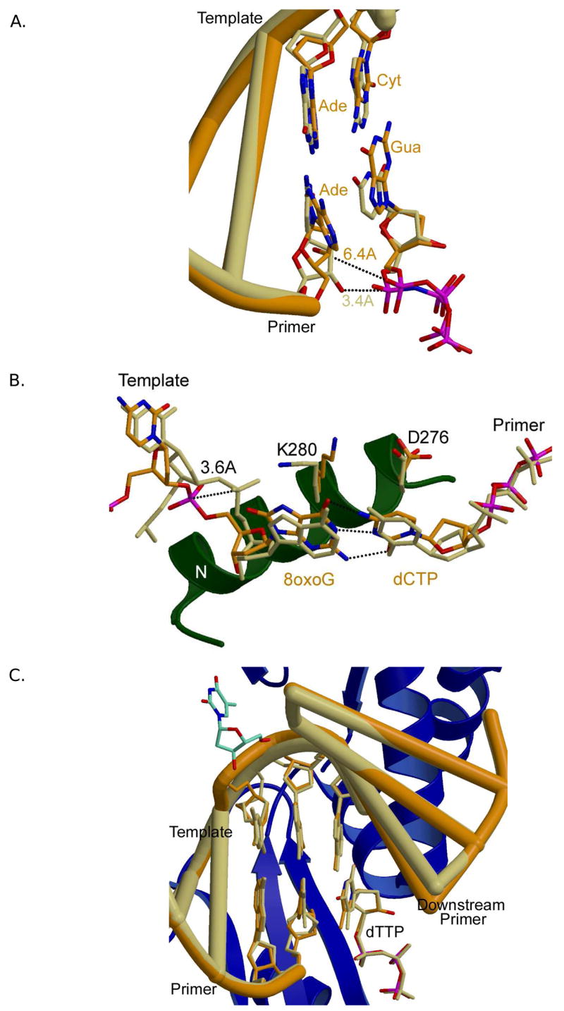
Accommodating distorted DNA substrates. (A) Superposition of a gapped substrate with an A-A mismatch at the primer terminus (PDB code 1ZJN, orange) and a normal gapped DNA substrate (PDB code 2FMS, khaki). The identities of the primer terminal and nascent base pairs are indicated (normal ternary in khaki, and mismatched ternary in orange). Identities of the DNA bases in the mismatch structure are shown to the right of each base (orange). (B) Superposition of a normal gapped DNA substrate (PDB code 2FMS, khaki) with canonical G-C base pair and a gapped DNA substrate containing an 8oxodG at the templating position (PDB code 1MQ3, orange), paired with incoming dCTP. (C) Pol λ accommodating an extra base. Superposition of a normal ternary complex of Pol λ (PDB code 1XSN; DNA, orange) with the frameshift intermediate (PDB code 2BCV; DNA khaki). Secondary structural elements for the frameshift structure are shown as blue ribbons. The extrahelical nucleotide is depicted in cyan. β-strand 8 harbors the K544 residue proximal to the extrahelical nucleotide, near the top of the panel.
References
-
- Bebenek K, Kunkel TA. Functions of DNA Polymerases. Adv Protein Chem. 2004;69:137–165. - PubMed
-
- Matsumoto Y, Kim K. Excision of deoxyribose phosphate residues by DNA polymerase beta during DNA repair. Science. 1995;269:699–702. - PubMed
-
- Garcia-Diaz M, Bebenek K, Kunkel TA, Blanco L. Identification of an intrinsic 5′-deoxyribose-5-phosphate lyase activity in human DNA polymerase lambda: a possible role in base excision repair. J Biol Chem. 2001;276:34659–34663. - PubMed
-
- Srivastava DK, Berg BJ, Prasad R, Molina JT, Beard WA, Tomkinson AE, Wilson SH. Mammalian abasic site base excision repair. Identification of the reaction sequence and rate-determining steps. J Biol Chem. 1998;273:21203–21209. - PubMed
-
- Braithwaite EK, Prasad R, Shock DD, Hou EW, Beard WA, Wilson SH. DNA polymerase lambda mediates a back-up base excision repair activity in extracts of mouse embryonic fibroblasts. J Biol Chem. 2005;280:18469–18475. - PubMed
Publication types
MeSH terms
Substances
Grants and funding
LinkOut - more resources
Full Text Sources
Other Literature Sources
Research Materials
