. 2007 Jul;133(1):268-77.
doi: 10.1053/j.gastro.2007.04.031. Epub 2007 Apr 20.

Effector role of neonatal hepatic CD8+ lymphocytes in epithelial injury and autoimmunity in experimental biliary atresia

Affiliations

Effector role of neonatal hepatic CD8+ lymphocytes in epithelial injury and autoimmunity in experimental biliary atresia

Pranavkumar Shivakumar et al. Gastroenterology. 2007 Jul.

Abstract

Background & aims: Lymphocytes populate the livers of infants with biliary atresia, but it is unknown whether neonatal lymphocytes regulate pathogenesis of disease. Here, we investigate this question by examining the role of T lymphocytes in the destruction of extrahepatic bile ducts of neonatal mice using an experimental model of biliary atresia.

Methods: Inoculation of neonatal mice with rhesus rotavirus followed by multistaining flow cytometry to quantify expression of interferon-gamma by hepatic lymphocytes, and real-time polymerase chain reaction for mRNA expression of pro-inflammatory cytokines. This was followed by determining the consequences of antibody-mediated depletion of lymphocyte subtypes on the development of biliary obstruction, and coculture and cell transfer experiments to investigate the effector role of lymphocyte subtypes on neonatal biliary disease.

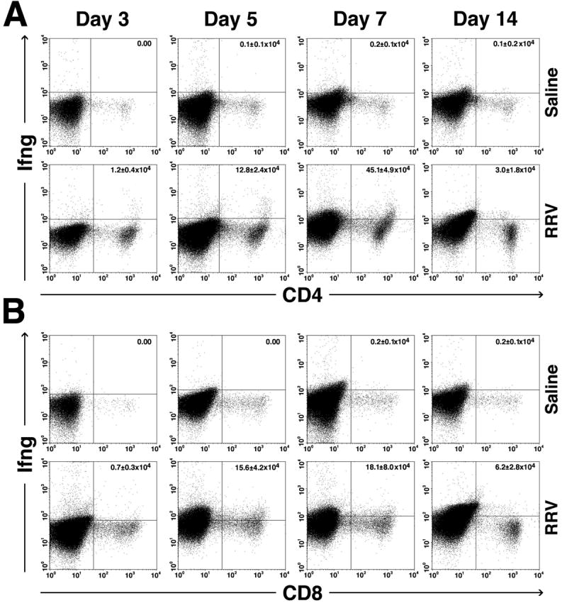
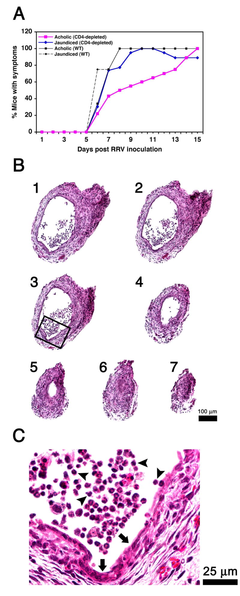
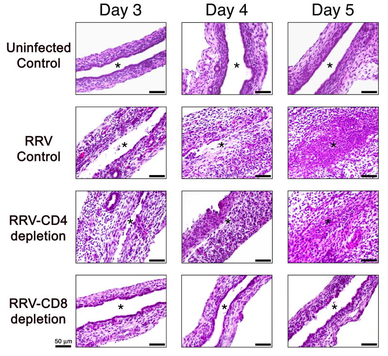
Results: Rotavirus infection results in overexpression of interferon-gamma by neonatal hepatic T cells. Among these cells, depletion of CD4(+) cells did not change the course of inflammatory injury and obstruction of neonatal bile ducts. In contrast, loss of CD8(+) cells remarkably suppressed duct injury, prevented luminal obstruction, and restored bile flow. Coculture experiments showed that rotavirus-primed, but not naïve, CD8(+) cells were cytotoxic to cholangiocytes. In adoptive transfer experiments, we found that primed CD8(+) cells preferentially homed to extrahepatic bile ducts of neonatal mice and invaded their epithelial lining.

Conclusions: Primed neonatal CD8(+) cells can activate a pro-inflammatory program, target diseased and healthy duct epithelium, and drive the phenotypic expression of biliary atresia, thus constituting a potential therapeutic target to halt disease progression.


Figures

Figure 1
Figure 1. Increased expression of Ifng by hepatic CD4+ and CD8+ cells after RRV challenge
Dot plots of flow cytometric analyses after surface and cytoplasmic staining of hepatic mononuclear cells with anti-CD4 or CD8 and Ifng antibodies show an increase in the number of CD4+ and CD8+ cells expressing Ifng as early as 3 days after RRV challenge, with peaks at the time of obstruction of extrahepatic bile ducts (7 days). Minimal to no dual staining is present in cells from saline-injected (control) mice. The numbers in right upper quadrants represent the mean ± standard deviation for the total number of hepatic CD4+ or CD8+ cells expressing Ifng. Each group contains N=3 mice per time point; P<0.05 when the RRV group is compared to the saline group for each time point.
Figure 2
Figure 2. Loss of CD4+ cells does not modify the course of bile duct obstruction
The onset of jaundice and acholic stools following RRV challenge in Balb/c mice was mildly delayed but the final appearance of symptoms in all mice was not altered by the antibody-mediated depletion of CD4+ cells (A). In B, successive cross-sectional views of bile ducts of CD4-depleted mice show infiltration of duct lumen by inflammatory cells (sections 1-4) and luminal obstruction (sections 5-7). The rectangle in section #3 is magnified in C, depicting epithelial injury and loss (arrows) and adjacent inflammatory cells (arrowheads). Hematoxylin/eosin stain; N=22 for WT mice infected with RRV; N=24 for CD4-depleted mice infected with RRV; sections are numbered from 1 to 7 to denote direction from the liver to the duodenum; WT = wild-type mice not injected with antibodies.
Figure 3
Figure 3. Loss of CD8+ cells improves symptoms and prevents luminal obstruction of bile ducts
RRV challenge of newborn mice induced the timely onset of jaundice and acholic stools in experimental and control groups. However, loss of CD8+ cells resulted in the resolution of symptoms in most mice by day 12 post infection (A). In B, successive cross-sectional views of bile ducts of CD8-depleted mice show infiltration of the subendothelial space by inflammatory cells (pericholangitis), but lack of intraluminal inflammation. The rectangle in section #1 is magnified in C, showing subepithelial inflammation (arrowheads) and intact epithelium (arrows). Hematoxylin/eosin stain; N=22 for WT mice infected with RRV; N=21 for CD8+ depleted mice infected with RRV; numbers 1 to 6 to denote direction from the liver to the duodenum; WT = wild-type mice not injected with antibodies.
Figure 4
Figure 4. Depletion of CD4+ or CD8+ cells does not modify expression of Ifng by lymphocytes in response to RRV
Flow cytometric analysis depicting an increased number of Ifng-expressing CD4+ or CD8+ cells in livers of mice injected with RRV or saline in the first day of life. All mice were subjected to antibody depletion of either CD4+ (red) or CD8+ (blue) cells; N=3 mice per group/time point; *P<0.05 when individual RRV-injected group is compared to saline-injected control.
Figure 5
Figure 5. Loss of CD8+ cells decreases injury to bile duct epithelium
Hematoxylin/eosin staining of longitudinal sections of extrahepatic bile ducts from uninfected controls and RRV challenged mice with or without depletion of CD4+ or CD8+ cells. An intact duct epithelium is present on days 3-5 in normal mice. In CD8-depleted mice, the epithelium also appears normal at day 3 and 4 and displays a mild focal epithelial injury at 5 days. In contrast, bile ducts of RRV-infected controls or CD4-depleted mice show extensive duct injury and obstruction at 4-5 days. Asterisk denotes the bile duct lumen.
Figure 6
Figure 6. RRV-primed hepatic CD8+ T cells induce cholangiocyte lysis
51Cr release assays showing increased lysis of a murine cholangiocyte cell line (mCL) by CD8+ T cells isolated from newborn mice 7 days after RRV challenge, which increases several fold above baseline levels of CD8+ T cells from saline-injected (control) mice. The specificity of the findings is shown by the lack of lysis of the 67-NR breast cancer cell line by CD8+ or CD4+ cells and by the inability of RRV-primed CD4+ cells to promote lysis of mCL cells. Ratio represents mCL or 67-NR cells (as target cells) to CD8+ cells (as effector cells). Results are representative of two independent experiments, with hepatic lymphocytes obtained from a pool of 15-20 livers for each experiment.
Figure 7
Figure 7. RRV-primed hepatic CD8+T cells have tropism to the biliary epithelium
Immunostaining of extrahepatic bile ducts with anti-CD3 and anti-cytokeratin antibodies shows a greater number of CD3+ cells in the subepithelial space (A) and within the epithelium (B) in mice transplanted with RRV-primed hepatic CD8+ cells. C depicts the red/pink staining in CD3+ cells and green staining in cytokeratin+ duct epithelium. C1 is from wild-type mice infected with RRV; C2 is from Rag2-/- mice transplanted with RRV-naïve hepatic CD8+ cells; and C3 and 4 are from Rag2-/- mice transplanted with RRV-primed CD8+ cells. Magnification × 6000; *denotes P<0.05 when RRV-CD4+ or RRV-CD8+ cells are compared to RRV-naïve CD8+ controls; N=4 mice per group.

References

    1. Garcia AM, Fadel SA, Cao S, Sarzotti M. T cell immunity in neonates. Immunol Res. 2000;22:177–90. - PubMed
    1. Adkins B. Development of neonatal Th1/Th2 function. Int Rev Immunol. 2000;19:157–71. - PubMed
    1. Brazolot Millan CL, Weeratna R, Krieg AM, Siegrist CA, Davis HL. CpG DNA can induce strong Th1 humoral and cell-mediated immune responses against hepatitis B surface antigen in young mice. Proc Natl Acad Sci U S A. 1998;95:15553–8. - PMC - PubMed
    1. Bot A, Bot S, Bona C. Enhanced protection against influenza virus of mice immunized as newborns with a mixture of plasmids expressing hemagglutinin and nucleoprotein. Vaccine. 1998;16:1675–82. - PubMed
    1. Hassett DE, Zhang J, Slifka M, Whitton JL. Immune responses following neonatal DNA vaccination are long-lived, abundant, and qualitatively similar to those induced by conventional immunization. J Virol. 2000;74:2620–7. - PMC - PubMed

Publication types

MeSH terms