. 2007 Jul;133(1):278-87.
doi: 10.1053/j.gastro.2007.04.032. Epub 2007 Apr 20.

Oligoclonal expansions of CD4+ and CD8+ T-cells in the target organ of patients with biliary atresia

Affiliations

Oligoclonal expansions of CD4+ and CD8+ T-cells in the target organ of patients with biliary atresia

Cara L Mack et al. Gastroenterology. 2007 Jul.

Abstract

Background & aims: Biliary atresia is an inflammatory, fibrosclerosing neonatal cholangiopathy, characterized by a periductal infiltrate composed of CD4(+) and CD8(+) T cells. The pathogenesis of this disease has been proposed to involve a virus-induced, subsequent autoreactive T cell-mediated bile duct injury. Antigen-specific T-cell immunity involves clonal expansion of T cells expressing similar T-cell receptor (TCR) variable regions of the beta-chain (Vbeta). We hypothesized that the T cells in biliary atresia tissue expressed related TCRs, suggesting that the expansion was in direct response to antigenic stimulation.

Methods: The TCR Vbeta repertoire of T cells from the liver, extrahepatic bile duct remnants, and peripheral blood of biliary atresia and other cholestatic disease controls were characterized by fluorescent-activated cell sorter analysis, and TCR junctional region nucleotide sequencing was performed on expanded TCR Vbeta regions to confirm oligoclonality.

Results: FACS analysis revealed Vbeta subset expansions of CD4(+) and CD8(+) T cells from the liver or bile duct remnant in all patients with biliary atresia and only 1 control. The CD4(+) TCR expansions were limited to Vbeta3, -5, -9, and -12 T-cell subsets and the CD8(+) TCR Vbeta expansions were predominantly Vbeta20. Each Vbeta subset expansion was composed of oligoclonal populations of T cells.

Conclusions: Biliary atresia is associated with oligoclonal expansions of CD4(+) and CD8(+) T cells within liver and extrahepatic bile duct remnant tissues, indicating the presence of activated T cells reacting to specific antigenic stimulation. Future studies entail identifying the specific antigen(s) responsible for T-cell activation and bile duct injury.


Figures

Figure 1
Figure 1
The sequence of events in recombination and gene expression of the TCR β-chain. Expressed TCR β-chain (TCRB) genes are generated from the rearrangement of variable (V) to diversity (D) to junctional (J) region gene segments. Further diversification is generated from the presence of random nucleotide additions and deletions at the V-to-D and D-to-J joining points. Shown here is an example of the TCRB region encoded by the selection of exons Vβ1, Dβ1, the second exon in the Jβ1 cluster, and the constant region Cβ1. This process makes the potential TCR repertoire enormous with greater than 107 sequence possibilities.
Figure 2
Figure 2
Representative histology of liver and extrahepatic bile duct remnant from BA and control tissue. Marked inflammation was observed within the portal tracts (A) and extra-hepatic bile duct remnant (B) of BA patients. Intense portal tract inflammation was also seen within the liver of control subjects (C, TPN-related cholestasis liver), while mild inflammation was present in extrahepatic bile duct tissue from control subjects (D, choledochal cyst).
Figure 3
Figure 3
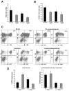
High yield of immune cells from BA tissue with a predominance of CD8+ T cells in liver tissue and CD4+ T cells in the extrahepatic duct remnant. (A) After 2 weeks in culture with IL-2, total cell yield was determined with a hemocytometer. Greater numbers of immune cells were obtained from BA livers and extrahepatic duct remnants compared to controls. (B) Percentage of total cells isolated in culture that were positive for the T-cell marker CD3. Significantly more CD3+ T cells were present in the liver from BA patients compared to controls (*P < .05). (C) Representative FACS analysis density plots of isolated T cells from BA specimens and controls, gated on all cells. Shown is a representative percentage of CD3+ T cells that were CD4 or CD8 positive in each group. (D) Summary of cell surface expression of CD3, CD4, and CD8. Significantly greater percentages of CD3+CD8+ T cells and smaller percentages of CD3+CD4+ T cells were detected from BA livers compared with liver controls (*P < .05).
Figure 4
Figure 4
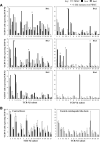
Expansion of CD4+ T cells with limited Vβ repertoire in BA livers and extrahepatic duct remnants. TCR Vβ repertoire of cultured CD4+ T cells from liver (shaded black), bile duct tissue (shaded gray), and PBMCs (white) in BA patients (A) and controls (B). Asterisks denote expanded TCR Vβ regions (>3-fold increase over PBMCs) in individual patients. All BA tissue contained 1 or 2 CD4+ TCR Vβ expansions. Shown in B are the mean ± SEM percent of CD4+ T cells expressing each of the Vβ subtypes from control tissues. Only 1 control tissue (choledochal cyst bile duct) had a significant CD4+ TCR Vβ expansion (Vβ17).
Figure 5
Figure 5
TCRB junctional region amino acid sequences expressed in CD4+ T cells from liver and extrahepatic duct remnants of BA patients. Dominant TCRB sequences are shown here. For each T-cell clone the entire TCRB junctional region is shown, extending from the 5′ end of the selected TCRBV family gene, including the highly rearranged nBDn gene segment, and ending at the selected BJ gene segment. The column TCRBJ denotes for each clone for which a BJ gene family member was selected during genetic rearrangement. The number of identical sequences is shown over the total number of sequences analyzed for a given sample and is depicted as percentage of frequency.
Figure 6
Figure 6
Striking expansion of CD8+ TCR Vβ20 in BA liver and extra-hepatic duct remnants. TCR Vβ repertoire of cultured CD8+ T cells from livers (shaded black) and bile duct tissue (shaded gray) compared with autologous PBMCs (white) in BA patients (A) and controls (B). Asterisks denote the expanded TCR Vβ region (>3-fold increase over PBMCs) in individual patients. Four of 6 BA specimens revealed striking expansions of CD8+ TCR Vβ20 within the liver and ductal remnants compared with autologous PBMCs. Shown in B are the mean ± SEM percent of CD8+ T cells expressing each of the Vβ subsets from control tissues. None of the controls contained CD8+ TCR Vβ expansions.
Figure 7
Figure 7
TCRB junctional region amino acid sequences expressed in CD8+ T cells expressing TCR Vβ20 from liver and extrahepatic duct remnants of BA patients. CD8+ TCR Vβ20 TCRB sequences found 3 times or more in any sample are shown here. For each T-cell clone the entire TCRB junctional region is shown. The column TCRBJ denotes for each clone for which a BJ gene family member was selected during genetic rearrangement. The number of identical sequences is shown over the total number of sequences analyzed for a given sample and is depicted as percentage frequency.

References

    1. Sokol RJ, Mack CL. Etiopathogenesis of biliary atresia. Semin Liver Dis. 2001;21:517–524. - PubMed
    1. Schreiber RA, Kleinman RE. Genetics, immunology and biliary atresia: an opening or a diversion? J Pediatr Gastroenterol Nutr. 1993;16:111–113. - PubMed
    1. Morecki R, Glaser JH, Cho S, Balistreri WF, Horwitz MS. Biliary atresia and reovirus type 3 infection. N Engl J Med. 1982;307:481–484. - PubMed
    1. Tyler KL, Sokol RJ, Oberhaus SM, Le M, Karrer FM, Narkewicz MR, Tyson RW, Murphy JR, Low R, Brown WR. Detection of reovirus RNA in hepatobiliary tissues from patients with extrahepatic biliary atresia and choledochal cysts. Hepatology. 1998;27:1475–1482. - PubMed
    1. Riepenhoff-Talty M, Gouvea V, Evans MJ, Svensson L, Hoffenberg E, Sokol RJ, Uhnoo I, Greenberg SJ, Schakel K, Zhaori G, Fitzgerald J, Chong S, el-Yousef M, Nemeth A, Brown M, Piccoli D, Hyams J, Ruffin D, Rossi T. Detection of group C rotavirus in infants with extrahepatic biliary atresia. J Infect Dis. 1996;174:8–15. - PubMed

Publication types

MeSH terms

Substances