. 2007 Oct 1;93(7):2542-52.
doi: 10.1529/biophysj.106.102426. Epub 2007 Jul 13.

Nuclear lamin A/C deficiency induces defects in cell mechanics, polarization, and migration

Affiliations

Nuclear lamin A/C deficiency induces defects in cell mechanics, polarization, and migration

Jerry S H Lee et al. Biophys J. .

Abstract

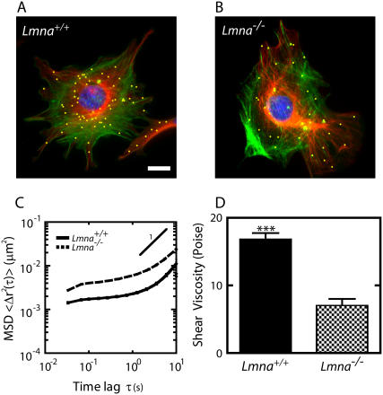
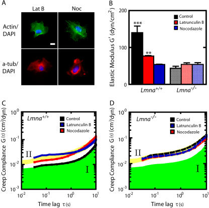
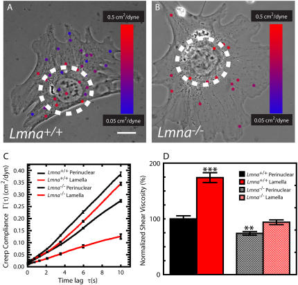
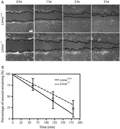
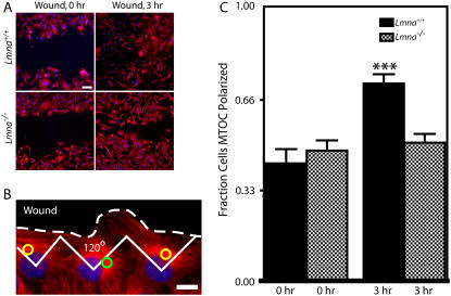
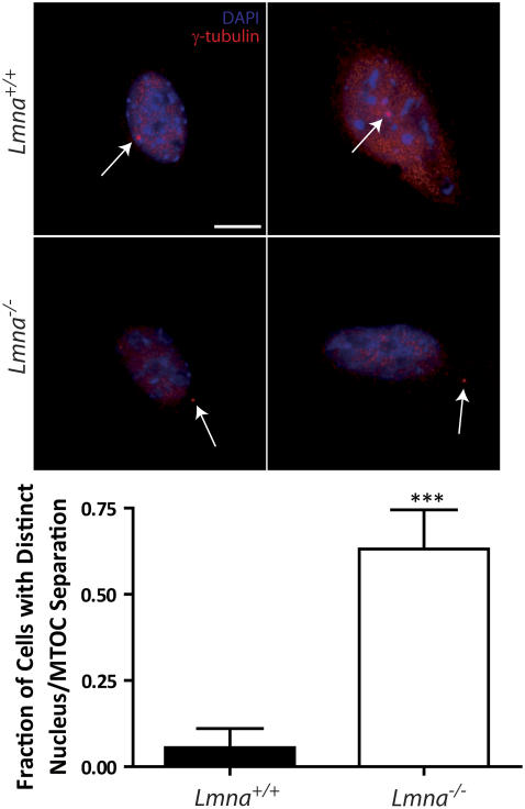
Lamin A/C is a major constituent of the nuclear lamina, a thin filamentous protein layer that lies beneath the nuclear envelope. Here we show that lamin A/C deficiency in mouse embryonic fibroblasts (Lmna(-/-) MEFs) diminishes the ability of these cells to polarize at the edge of a wound and significantly reduces cell migration speed into the wound. Moreover, lamin A/C deficiency induces significant separation of the microtubule organizing center (MTOC) from the nuclear envelope. Investigations using ballistic intracellular nanorheology reveal that lamin A/C deficiency also dramatically affects the micromechanical properties of the cytoplasm. Both the elasticity (stretchiness) and the viscosity (propensity of a material to flow) of the cytoplasm in Lmna(-/-) MEFs are significantly reduced. Disassembly of either the actin filament or microtubule networks in Lmna(+/+) MEFs results in decrease of cytoplasmic elasticity and viscosity down to levels found in Lmna(-/-) MEFs. Together these results show that both the mechanical properties of the cytoskeleton and cytoskeleton-based processes, including cell motility, coupled MTOC and nucleus dynamics, and cell polarization, depend critically on the integrity of the nuclear lamina, which suggest the existence of a functional mechanical connection between the nucleus and the cytoskeleton. These results also suggest that cell polarization during cell migration requires tight mechanical coupling between MTOC and nucleus, which is mediated by lamin A/C.


Figures

FIGURE 1
FIGURE 1
Altered mechanical properties of the cytoplasm in MEFs lacking lamin A/C. (A and B) Immunofluorescence micrographs of the actin filament (green) and microtubule (red) networks in Lmna+/+ MEFs (A) and Lmna−/− MEFs (B), overlaid with fluorescence micrographs of ballistically injected 100-nm diameter nanoparticles (yellow). Cells were fixed and actin, microtubule, and nuclear DNA were stained using Alexa-488 phallodin, α-tubulin/Alexa568, and DAPI, respectively. Nanoparticles were enlarged for ease of visualization. Bar, 20 μm. (C) Average mean-squared displacements (MSD) of nanoparticles imbedded in the cytoplasm of Lmna+/+ MEFs (solid line) and Lmna−/− MEFs (dashed line) (n > 35 cells each). A higher value of MSD indicates larger movements of the particles, which indicates a softer cytoplasm. (D) Average shear viscosity of the cytoplasm of Lmna+/+ MEFs (left) and Lmna−/− MEFs (right) calculated from the MSDs (n > 35 cells each).
FIGURE 2
FIGURE 2
Mechanical behavior of Lmna+/+ and Lmna−/− MEFs following actin filament or microtubule disassembly. (A) Immunofluorescence micrographs of the actin (green) and microtubule (red) networks in Lmna+/+ and Lmna−/− cells treated with either latrunculin B or nocadozole. Cells were fixed and actin, microtubule, and nuclear DNA were stained using Alexa-488 phallodin, α-tubulin/Alexa568, and DAPI, respectively. Bar, 20 μm. (B) Average elasticity of the cytoplasm of control and drug-treated Lmna+/+ (left columns) and Lmna−/− (right columns) MEFs (n > 25 cells were assayed for each condition). (C and D) Average creep compliance of the cytoplasm of control and drug-treated Lmna+/+ MEFs (C) and Lmna−/− MEFs (D). At least 25 cells were assayed for each condition. For ease of comparison, the green area indicates the compliance of control cells (regime I) and the yellow area indicates the compliance of the knockout cells (regime II).
FIGURE 3
FIGURE 3
Effect of lamin A/C deficiency on the mechanical properties of the perinuclear region and the lamella. (A and B) “Heat” maps of the local mechanical properties of MEFs. Phase contrast micrographs of Lmna+/+ MEF (A) and Lmna−/− MEFs (B) superimposed with fluorescence micrographs of the probe nanoparticles embedded in their cytoplasm. The circles are 30 μm in diameter and centered on the nuclei centers; they delineate the perinuclear region from the lamella. Nanoparticles are color-coded according to the local compliance of the cytoplasm in the vicinity of each nanoparticle. Blue corresponds to stiff regions and red to soft regions of the cell. The size of the nanoparticles is augmented for ease of view. Bar, 10 μm. (C) Average creep compliance of the perinuclear region and lamella of Lmna+/+ MEFs and Lmna−/− MEFs. At least 25 cells were assayed for type of cell. Linear scales are used for compliance and time lag to highlight differences. (D) Average shear viscosity in the perinuclear region and the lamella of Lmna+/+ MEFs and Lmna−/− MEFs. Shear viscosity values were normalized with the shear viscosity of the perinuclear region of Lmna+/+ MEFs. At least 25 cells were assayed for each type of cell.
FIGURE 4
FIGURE 4
Diminished speed of migration of MEFs lacking lamin A/C during wound healing. (A) Phase contrast micrographs of Lmna+/+ MEFs (top) and Lmna−/− MEFs (bottom) immediately after wounding, and 1, 2, and 3 h after wounding. The edges of the wounds are traced as guides to the eye. Bar, 100 μm. (B) Fraction of the wound that remains uncovered by the crawling cells as a function of time for Lmna+/+ MEFs (bottom) and Lmna−/− MEFs (top) (n = 7 for each type of cell).
FIGURE 5
FIGURE 5
Impaired MTOC repositioning in MEFs lacking lamin A/C. (A) Lmna+/+ MEFs and Lmna−/− MEFs immediately after a wound and 3 h after the wound. Cells were fixed and microtubule and nuclear DNA were stained using α-tubulin/Alexa568 and DAPI, respectively. Bar, 100 μm. (B) Microtubule network organization in Lmna+/+ MEFs at the edge of the wound. Cells at the edge of the wound, which have their MTOC preferentially located within the front third facing the wound, are considered polarized (yellow circles); cells that have their MTOC located in the back two-thirds of the cell are considered nonpolarized (green circle). Bar, 20 μm. (C) Fractions of Lmna+/+ MEFs and Lmna−/− MEFs that had a polarized MTOC before the wound (0 h) and 3 h after wounding. For Lmna+/+ MEFs, n = 91 cells at 0 h and n = 175 cells at 3 h. For Lmna−/− MEFs, n = 183 cells at 0 h and n = 242 cells at 3 h.
FIGURE 6
FIGURE 6
Separation between MTOC and nucleus induced by lamin A/C deficiency. (Top) Immnufluorescence micrographs of Lmna+/+ MEFs and Lmna−/− MEFs showing the position of the MTOC marked by γ-tubulin (thin arrows) stained using γ-tubulin/Alexa568 and the nucleus delineated by DAPI staining of nuclear DNA. Top row, typical Lmna+/+ MEFs; bottom row, typical Lmna−/− MEFs. Bar, 20 μm. (Bottom) Fraction of cells with a distinct separation between MTOC and nucleus. A value of 0 was assigned to a cell if the MTOC touched or laid on top or bottom of the nucleus, whereas a value of 1 was assigned to a cell if the MTOC was clearly separated from the nucleus. At least 20 cells were assayed for each type of cell.

References

    1. Hetzer, M. W., T. C. Walther, and I. W. Mattaj. 2005. Pushing the envelope: structure, function, and dynamics of the nuclear periphery. Annu. Rev. Cell Dev. Biol. 21:347–380. - PubMed
    1. Hutchison, C. J. 2002. Lamins: building blocks or regulators of gene expression? Nat. Rev. Mol. Cell Biol. 3:848–858. - PubMed
    1. Stuurman, N., S. Heins, and U. Aebi. 1998. Nuclear lamins: their structure, assembly, and interactions. J. Struct. Biol. 122:42–66. - PubMed
    1. Elbashir, S. M., J. Harborth, W. Lendeckel, A. Yalcin, K. Weber, and T. Tuschl. 2001. Duplexes of 21-nucleotide RNAs mediate RNA interference in cultured mammalian cells. Nature. 411:494–498. - PubMed
    1. Vergnes, L., M. Peterfy, M. O. Bergo, S. G. Young, and K. Reue. 2004. Lamin B1 is required for mouse development and nuclear integrity. Proc. Natl. Acad. Sci. USA. 101:10428–10433. - PMC - PubMed

Publication types