Review
. 2007 Aug;1767(8):1007-31.
doi: 10.1016/j.bbabio.2007.05.008. Epub 2007 Jun 2.

The role of mitochondria in protection of the heart by preconditioning

Affiliations
Review

The role of mitochondria in protection of the heart by preconditioning

Andrew P Halestrap et al. Biochim Biophys Acta. 2007 Aug.

Abstract

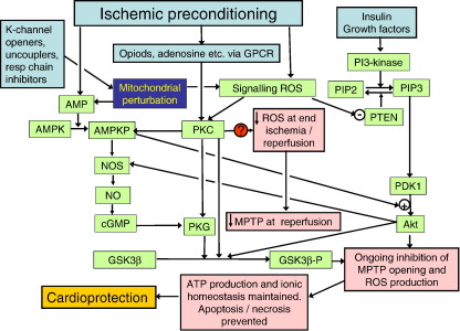
A prolonged period of ischaemia followed by reperfusion irreversibly damages the heart. Such reperfusion injury (RI) involves opening of the mitochondrial permeability transition pore (MPTP) under the conditions of calcium overload and oxidative stress that accompany reperfusion. Protection from MPTP opening and hence RI can be mediated by ischaemic preconditioning (IP) where the prolonged ischaemic period is preceded by one or more brief (2-5 min) cycles of ischaemia and reperfusion. Following a brief overview of the molecular characterisation and regulation of the MPTP, the proposed mechanisms by which IP reduces pore opening are reviewed including the potential roles for reactive oxygen species (ROS), protein kinase cascades, and mitochondrial potassium channels. It is proposed that IP-mediated inhibition of MPTP opening at reperfusion does not involve direct phosphorylation of mitochondrial proteins, but rather reflects diminished oxidative stress during prolonged ischaemia and reperfusion. This causes less oxidation of critical thiol groups on the MPTP that are known to sensitise pore opening to calcium. The mechanisms by which ROS levels are decreased in the IP hearts during prolonged ischaemia and reperfusion are not known, but appear to require activation of protein kinase Cepsilon, either by receptor-mediated events or through transient increases in ROS during the IP protocol. Other signalling pathways may show cross-talk with this primary mechanism, but we suggest that a role for mitochondrial potassium channels is unlikely. The evidence for their activity in isolated mitochondria and cardiac myocytes is reviewed and the lack of specificity of the pharmacological agents used to implicate them in IP is noted. Some K(+) channel openers uncouple mitochondria and others inhibit respiratory chain complexes, and their ability to produce ROS and precondition hearts is mimicked by bona fide uncouplers and respiratory chain inhibitors. IP may also provide continuing protection during reperfusion by preventing a cascade of MPTP-induced ROS production followed by further MPTP opening. This phase of protection may involve survival kinase pathways such as Akt and glycogen synthase kinase 3 (GSK3) either increasing ROS removal or reducing mitochondrial ROS production.


Figures

Fig. 1
Fig. 1
Preconditioning is not accompanied by translocation of protein kinases to the mitochondria. Isolated rat hearts were preconditioned using two cycles of 5 min ischaemia interspersed with 5 min reperfusion as described previously or treated for 10 min with 50 μM diazoxide or 200 nM phorbol-12-myristate-13-acetate (Phorbol ester). Five minutes after the second brief ischemic period or at the equivalent time for control and drug-treated hearts, mitochondria rapidly isolated in the presence of buffer containing proteases and phosphatase inhibitors as described in where further details of the Methodology are given. In Panel A samples of the post-mitochondrial supernatant (Cyt) and the mitochondria before (Crd) and after (Mit) removal of contaminating membranes by Percoll density gradient centrifugation were separated by SDS-PAGE and Western blotting performed using antibodies against PKCα, PKCε, MCT1 (as a sarcolemmal marker) and ANT (as a mitochondrial marker). In Panel B similar experiments were performed but in this case crude samples of crude homogenate (after clarification by centrifugation at 2000×g for 5 min) were included (Hom) as were samples of the membrane fraction (Slm—primarily sarcolemma) removed by Percoll density gradient centrifugation. Western blotting was performed using antibodies against total and phosphorylated forms of AMPK, GSK3β and Akt, as well as MCT1, ANT and glyceraldehyde-3-phosphate dehydrogenase (GAPDH) as markers for sarcolemma, mitochondria and cytosol. Data shown are previously unpublished data of the authors.
Fig. 2
Fig. 2
Preconditioning is not accompanied by phosphorylation of integral mitochondrial proteins. Density gradient purified mitochondria were isolated from control and preconditioned rat hearts 5 min after the preconditioning protocol as described in Fig. 1. Mitochondrial proteins were separated by 2D electrophoresis in the Proteomics Facility of the University of Bristol and gels stained with ProQDiamond to reveal phosphorylated proteins (red) followed by sypro-Ruby to stain all proteins (green). The fluorescent images were scanned after each staining procedure and the images overlayed. The predominant phosphoprotein at about 50 kDa and pI 6.5 were shown to be multiply phosphorylated forms of pyruvate dehydrogenase and branch-chain keto acid dehydrogenase that are known to be the dominant phosphoproteins in heart mitochondria .
Fig. 3
Fig. 3
No evidence for mitochondrial matrix volume changes induced by openers and blockers of the putative mitochondrial KATP channel. Data of Panels A and B are modified from where further details may be found. Panel A show the light scattering changes of energised rat heart mitochondria induced by the addition where indicated of 0.2 mM ATP, 5 μM carboxyatractyloside (CAT), 0.2 and 0.5 nM valinomycin (Val) and 50 μM 5HD, diazoxide or glibencamide. Where ATP or 5HD are present from the start this is indicated on the left of the trace. Panel B shows the corresponding matrix volume changes induced by valinomycin or ATP as measured using 3H2O and [14C]-sucrose. Panel C represents previously unpublished data that compares light scattering changes of energised mitochondria that are induced by ATP and CAT in KCl and tetraethylammonium (TEA) media. The latter was used by Garlid and colleagues (see [165]) as a potassium free medium to establish whether these changes are independent of K+ movements across the inner mitochondrial membrane.
Fig. 4
Fig. 4
Connexin 43 does not translocate to mitochondria following preconditioning. Percoll purified mitochondria were isolated from control and IP hearts in buffer containing protease and phosphatase inhibitors as described for Figs. 1 and2. A total particulate and cytosol fraction were also prepared by centrifugation for 45 min at 150,000×g. For Panel A, samples were subject to SDS-PAGE and Western blotting using the antibodies indicated. The inset blot shows greater MCT1 contamination in mitochondria prepared according to the method used by Boengler et al. . Panel B shows the data (mean ± S.E.M.) from 6 separate experiments in which the Cx43 and ANT blots were scanned and the Cx43 band intensity expressed as a ratio with respect to the ANT band intensity for mitochondrial and particulate fractions.
Fig. 5
Fig. 5
Suggested pathways by which IP, KATP channel openers and other factors that perturb mitochondrial function may lead to inhibition of MPTP opening during reperfusion. The proposed scheme is based on the evidence and reasoning presented in the text. Boxes shaded pastel blue represent the preconditioning stimuli, whilst pastel green represent components of potential signalling pathways and pastel pink the effects on the mitochondrial target. The blue box signifies the effects of any process that perturbs mitochondrial function such as through mild inhibition of respiratory chain complexes or uncoupling. The red circle and question mark that links PKC to diminished ROS levels at the end of ischaemia and during reperfusion target represent a critical step in preconditioning whose mechanism is unknown and requires elucidation.

Similar articles

Cited by

References

    1. Halestrap A.P., Kerr P.M., Javadov S., Woodfield K.Y. Elucidating the molecular mechanism of the permeability transition pore and its role in reperfusion injury of the heart. Biochim. Biophys. Acta. 1998;1366:79–94. - PubMed
    1. Suleiman M.S., Halestrap A.P., Griffiths E.J. Mitochondria: a target for myocardial protection. Pharmacol. Ther. 2001;89:29–46. - PubMed
    1. Halestrap A.P., Clarke S.J., Javadov S.A. Mitochondrial permeability transition pore opening during myocardial reperfusion—A target for cardioprotection. Cardiovasc. Res. 2004;61:372–385. - PubMed
    1. Downey J.M., Cohen M.V. Reducing infarct size in the setting of acute myocardial infarction. Prog. Cardiovasc. Dis. 2006;48:363–371. - PubMed
    1. Fliss H., Gattinger D. Apoptosis in ischemic and reperfused rat myocardium. Circ. Res. 1996;79:949–956. - PubMed

Publication types

MeSH terms

Substances