

Review
doi: 10.1016/j.mvr.2007.05.006.
Epub 2007 Jun 6.
In vitro assays of angiogenesis for assessment of angiogenic and anti-angiogenic agents
Affiliations
- PMID: 17631914
- PMCID: PMC2692317
- DOI: 10.1016/j.mvr.2007.05.006
Item in Clipboard
Review
In vitro assays of angiogenesis for assessment of angiogenic and anti-angiogenic agents
Microvasc Res.
2007 Sep-Nov.
Abstract
Blood vessels, either in insufficient numbers or in excess, contribute to the pathogenesis of many diseases. Agents that stimulate angiogenesis can improve blood flow in patients with ischemic diseases, whereas anti-angiogenic agents are used to treat disorders ranging from macular degeneration to cancer. In this review I describe in vitro assays that can be used to assess the activity of agents that affect angiogenesis. Means of quantifying endothelial cell matrix degradation, migration, proliferation, apoptosis and morphogenesis are discussed, as are embryoid body, aortic ring and metatarsal assays of vessel outgrowth. Strengths and limitations of these techniques are also addressed.
Figures

Endothelial cell functions involved in angiogenesis.
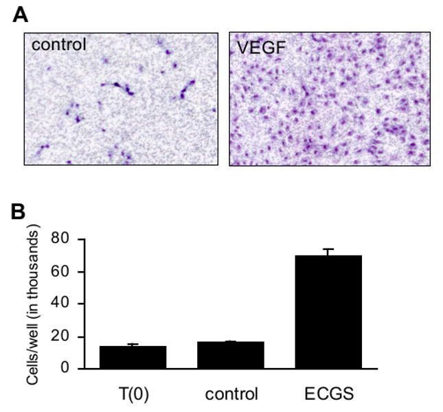
Cell culture assays assessing endothelial cell function. A) Cell migration as measured using a transwell assay. Starved HUVEC were plated onto a fibronectin-coated transwell with pores 3 μm pore diameter. Medium on the opposing side of the membrane lacked (left panel) or contained (right panel) recombinant VEGF at 10 ng/ml. After 18 hr, nonmigrated cells were removed with a cotton swab. Migrated cells were fixed and stained with hematoxylin and eosin, then photographed using a 5× objective. Unpublished images courtesy of Magali Saint-Geniez. B) Cell proliferation as measured by direct cell counting. HUVEC were treated with endothelial cell growth supplement and cultured for three days. The mean numbers of cells present in each well prior to the start of treatment (T(0)) and following three days of treatment (control, VEGF) are shown. Error bars indicate standard deviation. Unpublished data courtesy of Sandie Smith.
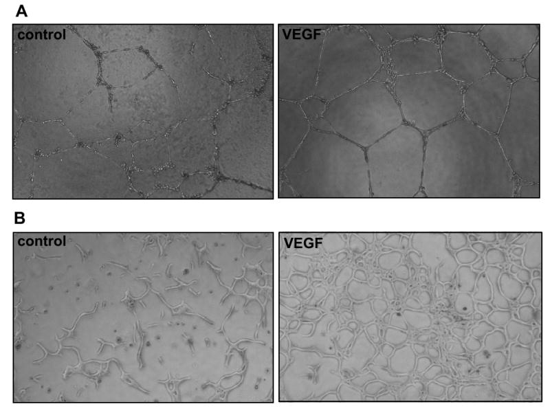
Endothelial cell morphogenesis. A) HUVEC plated on Matrigel. HUVEC were plated onto Matrigel pads and cultured in the absence (left panel) or presence (right panel) of VEGF (10 ng/ml). Capillary-like structures were photographed (10× objective) 20 hr after plating. B) Bovine aortic endothelial cells suspended in diluted Matrigel. The endothelial cells were suspended in a matrix containing 50% Matrigel and 50% culture medium. The following day, the medium was replaced with serum-free medium lacking (left panel) or containing (right panel) VEGF at 10 ng/ml. Capillary-like structures were photographed (10× objective) after three days of treatment. Unpublished images courtesy of Tony Walshe.
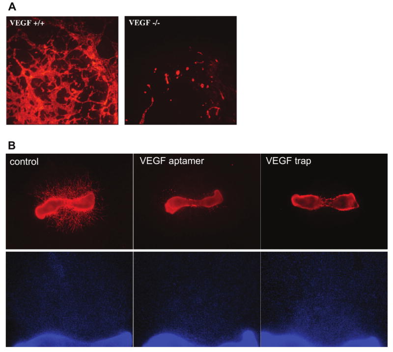
Embryoid body and mouse metatarsal models of angiogenesis. A) Vascular structures in embryoid bodies. Embryoid bodies were differentiated for 10 days from VEGF-deficient (left panel) or wild-type (right panel) mouse embryonic stem cells. The vascular structures were labeled using an antibody to PECAM and photographed at using a 5× objective. Unpublished images courtesy of Robyn Loureiro. B) Vessel outgrowth from mouse metatarsal bones. Metatarsal bones were isolated from wild type embryonic day 18 mouse embryos and cultured with control medium (left panels), an aptamer to VEGF (Ruckman et al., 1998; middle panels) or a soluble, truncated VEGF receptor (VEGF trap; Holash et al., 1992; right panels). Vessel outgrowth in the top panels is visualized by fluorescently labeling the endothelial cell marker PECAM and photographing using a 2× objective. The DAPI staining in the lower panels, photographed using a 4× objective, shows that the inhibitors do not prevent outgrowth of non-endothelial cells from the metatarsal bone. Unpublished images courtesy of Yin-Shan Ng.
References
-
- Ahmad S, Ahmad A, Schneider KB, White CW. Cholesterol interferes with the MTT assay in human epithelial-like (A549) and endothelial (HLMVE and HCAE) cells. Int J Toxicol. 2006;25:17–23. - PubMed
-
- Ashikari-Hada S, Habuchi H, Kariya Y, Kimata K. Heparin regulates vascular endothelial growth factor165-dependent mitogenic activity, tube formation, and its receptor phosphorylation of human endothelial cells. Comparison of the effects of heparin and modified heparins. J Biol Chem. 2005;280:31508–31515. - PubMed
-
- Auerbach R, Akhtar N, Lewis RL, Shinners BL. Angiogenesis assays: problems and pitfalls. Cancer Metastasis Rev. 2000;19:167–172. - PubMed
-
- Auerbach R, Lewis R, Shinners B, Kubai L, Akhtar N. Angiogenesis assays: a critical overview. Clin Chem. 2003;49:32–40. - PubMed
-
- Bautch VL, Stanford WL, Rapoport R, Russell S, Byrum RS, Futch TA. Blood island formation in attached cultures of murine embryonic stem cells. Dev Dyn. 1996;205:1–12. - PubMed
Publication types
MeSH terms
Substances
Grants and funding
LinkOut - more resources
Full Text Sources
Other Literature Sources
