. 2007 Oct 25;367(2):440-51.
doi: 10.1016/j.virol.2007.06.010. Epub 2007 Jul 12.

The interferon-induced expression of APOBEC3G in human blood-brain barrier exerts a potent intrinsic immunity to block HIV-1 entry to central nervous system

Affiliations

The interferon-induced expression of APOBEC3G in human blood-brain barrier exerts a potent intrinsic immunity to block HIV-1 entry to central nervous system

Elias G Argyris et al. Virology. .

Abstract

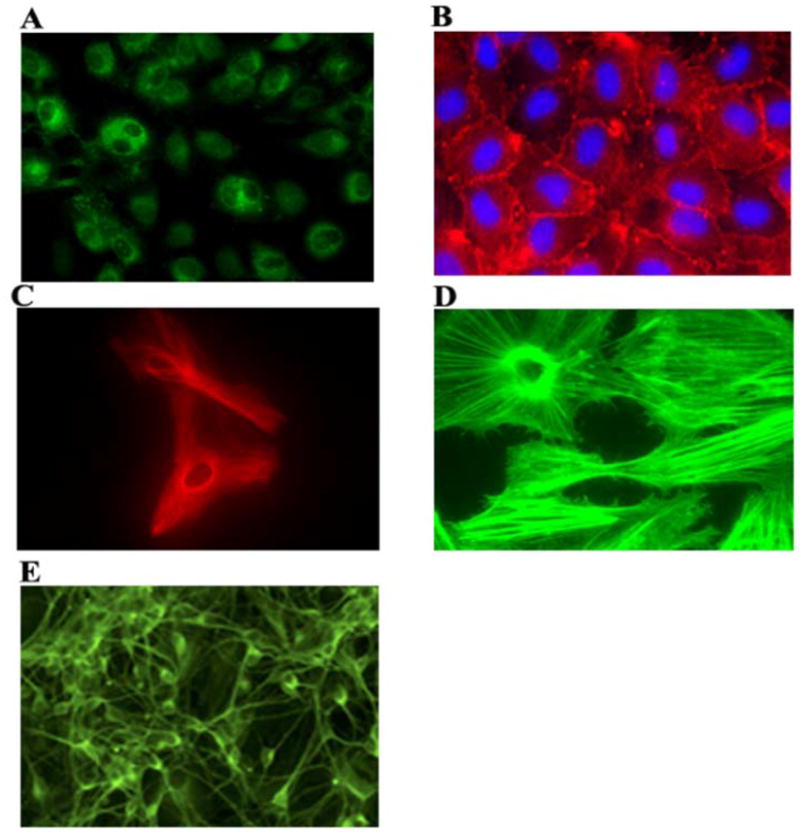
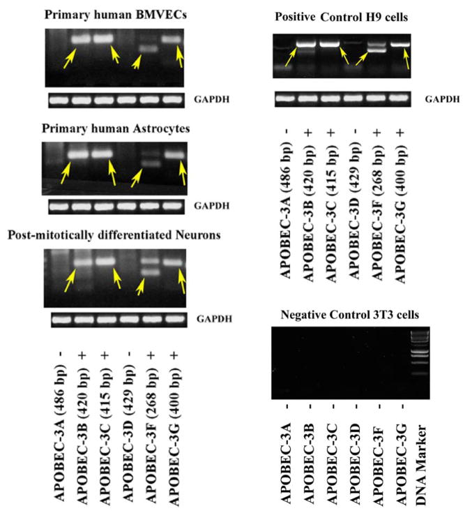
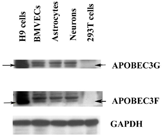
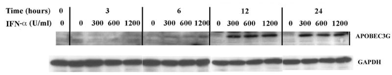
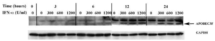
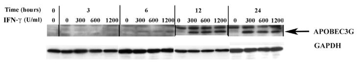
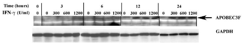
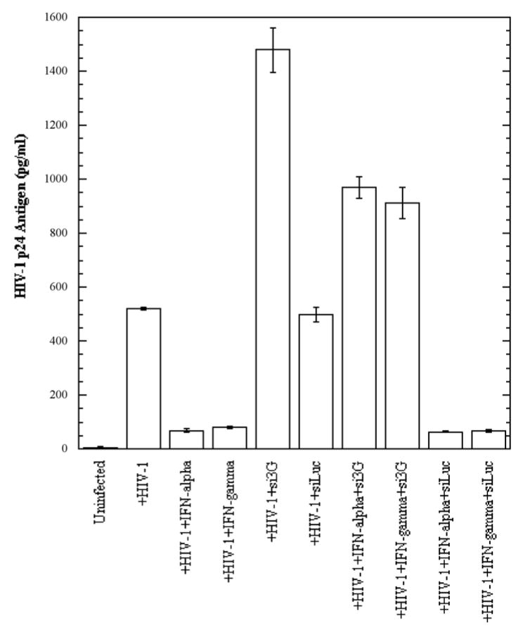
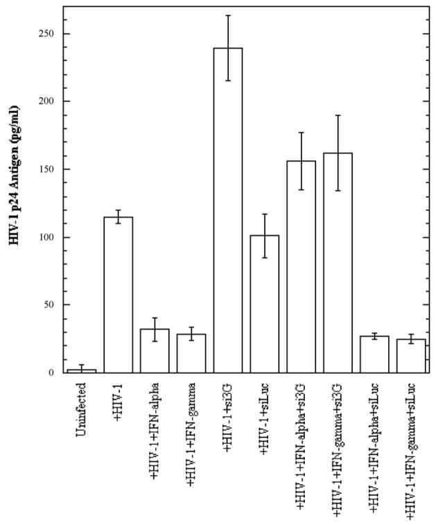
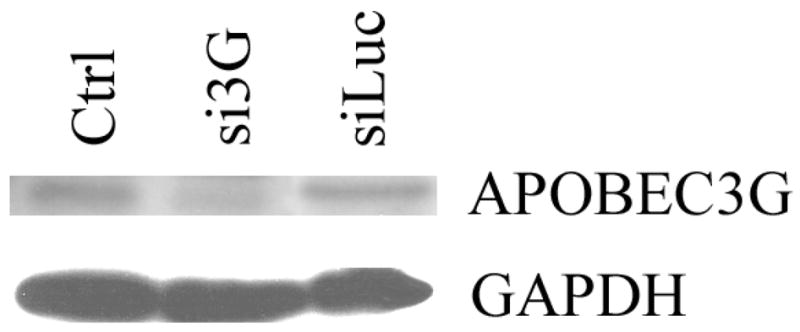
In the human genome, the APOBEC3 gene has expanded into a tandem array of genes termed APOBEC3A-H. Several members of this family have potent anti-HIV-1 activity. Here we demonstrate that APOBEC-3B/3C/3F and -3G are expressed in all major cellular components of the CNS. Moreover, we show that both interferon-alpha (IFN-alpha) and IFN-gamma significantly enhance the expression of APOBEC-3G/3F and drastically inhibit HIV-1 replication in primary human brain microvascular endothelial cells (BMVECs), the major component of blood-brain barrier (BBB). As the viral inhibition can be neutralized by APOBEC3G-specific siRNA, APOBEC3G plays a key role to mediate the anti-HIV-1 activity of IFN-alpha and/or IFN-gamma. Our findings suggest that, in addition to the restriction at viral entry level, the restriction from APOBEC3 family could account for the low-level replication of HIV-1 in BMVECs. The manipulation of IFN-APOBEC3 signaling pathway could be a potent therapeutic strategy to prevent HIV invasion to central nervous system (CNS).


Figures

Figure 1
Figure 1. Primary human CNS-based cell systems used in our studies
All CNS-based cell systems were cultured and maintained as described in Materials and Methods. Shown are: Primary human BMVECs expressing von Willebrand Factor (A), and ZO-1 (B). Primary human brain astrocytes expressing glial fibrillary acidic protein (C), and stained for cytoskeletal structural proteins (D). Post-mitotically differentiated neurons immunostained for MAP-2 (E).
Figure 2
Figure 2. Expression of APOBEC3 family proteins in human CNS-based cell cultures
(A). Reverse transcriptase (RT)-PCR was employed to detect the presence of APOBEC-3A/3B/3C/3D/3F and 3G mRNA isolated from primary human brain microvascular endothelial cells (BMVECs), astrocytes and differentiated post-mitotic mature neuronal cells which are key components of the blood-brain barrier (BBB) and the CNS, as well as control H9 cells. 3T3 cells served as negative control. Arrows indicate the specifically amplified APOBEC3 products: Apobec-3B, -3C, -3F and –3G were detected (“+” positive), while Apobec-3A and –3D were not detected (“−” negative). GAPDH served as a positive control. (B). Cell lysates from primary human BMVECs, astrocytes and differentiated post-mitotic neuronal cells, as well as H9 (positive control) and 293T cells (negative control) were analyzed via Western blot using rabbit polyclonal antibodies against APOBEC3G (~46 kD) and APOBEC3F (~46 kD). GAPDH (~36 kD) served as a positive control.
Figure 3
Figure 3. APOBEC3G and APOBEC3F expression is upregulated by IFN-α in human primary BMVECs
(A). Time course and dose-dependent studies on the upregulation of APOBEC3G by IFN-α. IFN-α (0, 300, 600 and 1200 U/ml) was added into cultures of primary human BMVECs. The intracellular expression of APOBEC3G protein was assessed by Western-blot analysis of cell lysate samples collected at various time points: 0-, 3-, 6-, 12- and 24-hrs post IFN-α treatment. (B). Time course and dose-dependent studies on the upregulation of APOBEC3F by IFN-α. IFN-α (0, 300, 600 and 1200 U/ml) was added into cultures of primary human BMVECs. The intracellular expression of APOBEC3F protein was assessed by Western-blot analysis of cell lysate samples collected at various time points: 0-, 3-, 6-, 12- and 24- hrs post IFN-α treatment.
Figure 4
Figure 4. IFN-γ also upregulates the expression of APOBEC3G and APOBEC3F in human primary BMVECs
(A). Time course and dose-dependent studies on the upregulation of AOBEC3G by IFN-γ. IFN-γ (0, 300, 600 and 1200 U/ml) was added into cultures of primary human BMVECs. The intracellular expression of APOBEC3G protein was assessed by Western-blot analysis of cell lysate samples collected at various time points: 0-, 3-, 6-, 12- and 24- hrs post IFN-γ treatment. (B). Time course and dose-dependent studies on the upregulation of APOBEC3F by IFN-γ. IFN-γ (0, 300, 600 and 1200 U/ml) was added into cultures of primary human BMVECs. The intracellular expression of APOBEC3F protein was assessed by Western-blot analysis of cell lysate samples collected at various time points: 0-, 3-, 6-, 12- and 24-hrs post IFN-γ treatment.
Figure 5
Figure 5. siRNA against APOBEC3G blocks IFN-α and IFN-γ anti-HIV-1 activity in human primary BMVECs
(A). Primary human BMVECs cultured at 50% confluence in 12-well plates were transfected with and without APOBEC3G-specific siRNA (si3G) or luciferase-specific siRNA (siLuc) (100 nmol/ml). After 72 hrs, the transfected or untransfected BMVECs were infected with HIV-1-VSV-GFP virus (20 ng of p24 equivalents) for 6 hrs. Simultaneously, the infected cells were treated with and without IFN-α or IFN-γ (300 U/ml) for 72 hrs. HIV-1 p24 antigen in cell lysates normalized for protein content was detected by ELISA at day three post-infection (3 d.p.i.). The data represent two independent experiments. (B). Primary human BMVECs cultured at 50% confluence in 12-well plates were transfected with or without si3G or siLuc (100 nmol/ml). After 72 hrs, the transfected or untransfected BMVECs were infected with the R5-tropic HIV-1 strain YU2 (20 ng of p24 equivalents) for 6 hrs. Simultaneously, the infected cells were treated with or without IFN-α or IFN-γ (300 U/ml) for 72 hrs. HIV-1 p24 antigen in cell lysates normalized for protein content was detected by ELISA at day three post-infection (3 d.p.i.). The data represent two independent experiments. (C). Western blot for APOBEC3G protein 6 days post-transfection of primary human BMVECs with DharmaFECT 1 transfection reagent (Ctrl), APOBEC3G-specific siRNA (si3G) or luciferase-specific siRNA (siLuc).

References

    1. Acheampong E, Parveen Z, Mengistu A, Ngoubilly N, Wigdahl B, Lossinsky AS, Pomerantz RJ, Mukhtar M. Cholesterol-depleting statin drugs protect postmitotically differentiated human neurons against ethanol- and human immunodeficiency virus type 1-induced oxidative stress in vitro. J Virol. 2007;81(3):1492–501. - PMC - PubMed
    1. Argyris EG, Acheampong E, Nunnari G, Mukhtar M, Williams KJ, Pomerantz RJ. Human immunodeficiency virus type 1 enters primary human brain microvascular endothelial cells by a mechanism involving cell surface proteoglycans independent of lipid rafts. J Virol. 2003;77(22):12140–51. - PMC - PubMed
    1. Bagasra O, Lavi E, Bobroski L, Khalili K, Pestaner JP, Tawadros R, Pomerantz RJ. Cellular reservoirs of HIV-1 in the central nervous system of infected individuals: identification by the combination of in situ polymerase chain reaction and immunohistochemistry. Aids. 1996;10(6):573–85. - PubMed
    1. Bell JE, Busuttil A, Ironside JW, Rebus S, Donaldson YK, Simmonds P, Peutherer JF. Human immunodeficiency virus and the brain: investigation of virus load and neuropathologic changes in pre-AIDS subjects. J Infect Dis. 1993;168(4):818–24. - PubMed
    1. Berglund O, Engman K, Ehrnst A, Andersson J, Lidman K, Akerlund B, Sonnerborg A, Strannegard O. Combined treatment of symptomatic human immunodeficiency virus type 1 infection with native interferon-alpha and zidovudine. J Infect Dis. 1991;163(4):710–5. - PubMed

Publication types

MeSH terms