. 2007 Oct 25;367(2):324-33.
doi: 10.1016/j.virol.2007.06.011. Epub 2007 Jul 13.

Iron chelators ICL670 and 311 inhibit HIV-1 transcription

Affiliations

Iron chelators ICL670 and 311 inhibit HIV-1 transcription

Zufan Debebe et al. Virology. .

Abstract

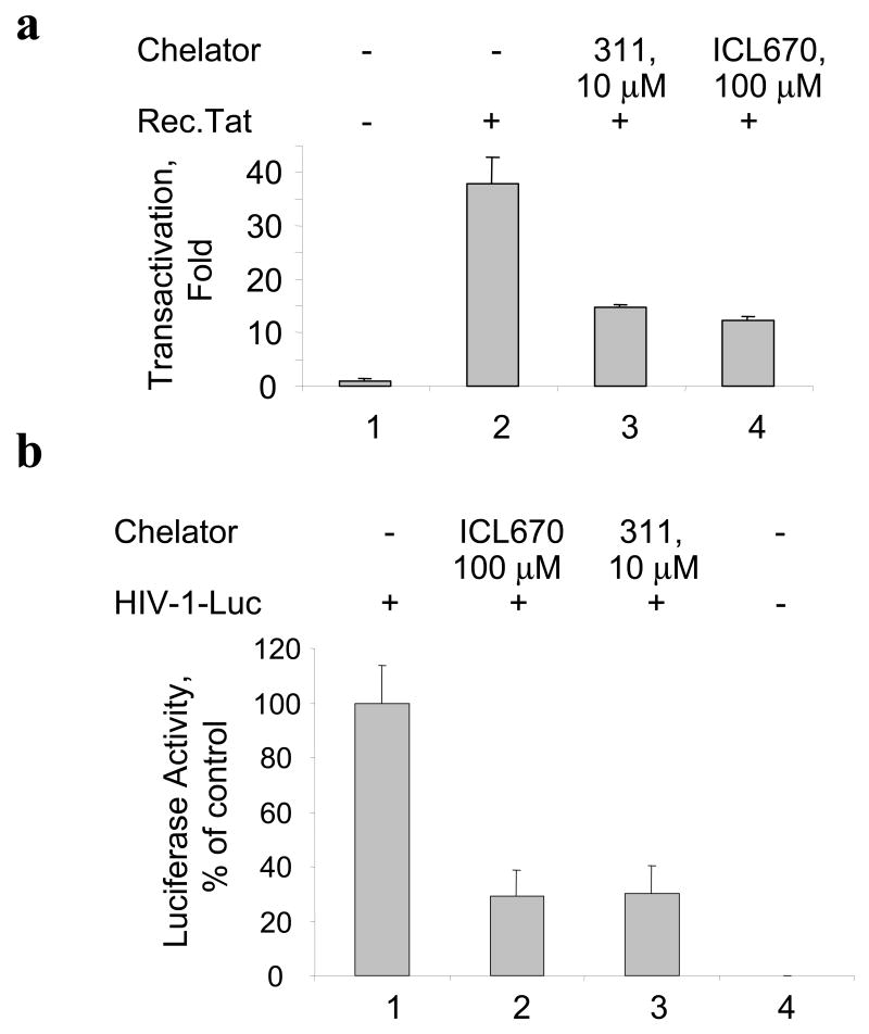
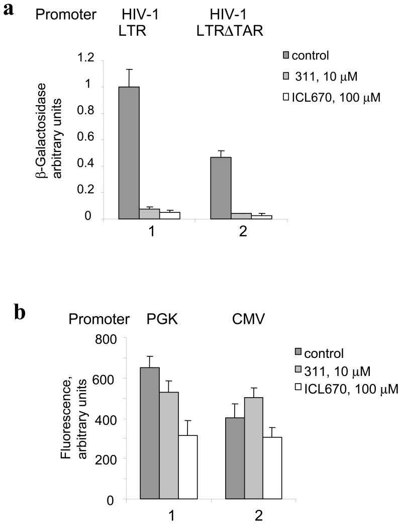
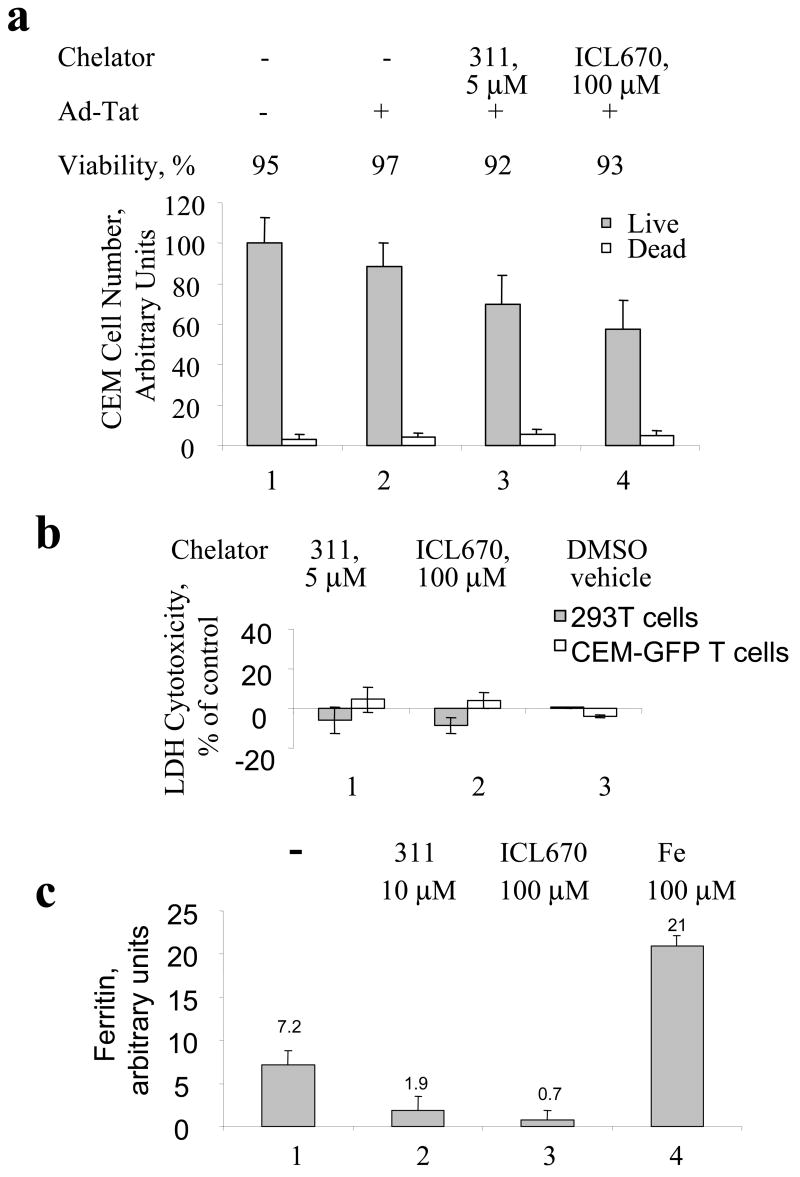
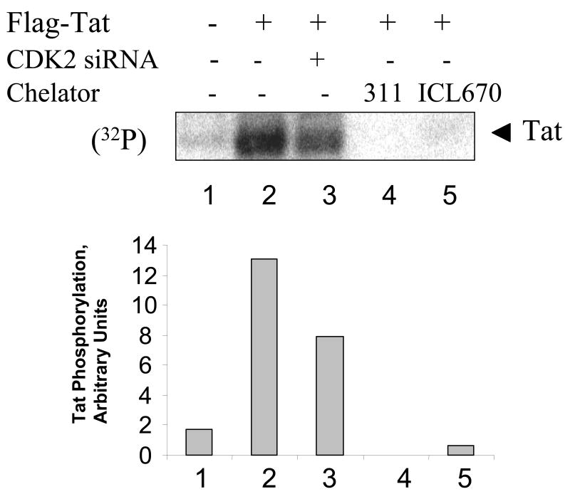
HIV-1 replication is induced by an excess of iron and iron chelation by desferrioxamine (DFO) inhibits viral replication by reducing proliferation of infected cells. Treatment of cells with DFO and 2-hydroxy-1-naphthylaldehyde isonicotinoyl hydrazone (311) inhibit expression of proteins that regulate cell-cycle progression, including cycle-dependent kinase 2 (CDK2). Our recent studies showed that CDK2 participates in HIV-1 transcription and viral replication suggesting that inhibition of CDK2 by iron chelators might also affect HIV-1 transcription. Here we evaluated the effect of a clinically approved orally effective iron chelator, 4-[3,5-bis-(hydroxyphenyl)-1,2,4-triazol-1-yl]-benzoic acid (ICL670) and 311 on HIV-1 transcription. Both ICL670 and 311 inhibited Tat-induced HIV-1 transcription in CEM-T cells, 293T and HeLa cells. Neither ICL670 nor 311 induced cytotoxicity at concentrations that inhibited HIV-1 transcription. The chelators decreased cellular activity of CDK2 and reduced HIV-1 Tat phosphorylation by CDK2. Neither ICL670A or 311 decreased CDK9 protein level but significantly reduced association of CDK9 with cyclin T1 and reduced phosphorylation of Ser-2 residues of RNA polymerase II C-terminal domain. In conclusion, our findings add to the evidence that iron chelators can inhibit HIV-1 transcription by deregulating CDK2 and CDK9. Further consideration should be given to the development of iron chelators for future anti-retroviral therapeutics.


Figures

Fig. 1
Fig. 1. Iron chelators 311 and ICL670 inhibit Tat-induced HIV-1 transcription in CEM T-cells
(a) CEM-GFP T-cells were grown in 96-well plate and infected with the indicated amounts of Ad-Tat or Ad-LacZ. At 24 h the cells were lysed and GFP fluorescence was measured on a luminescence spectrometer. (b) CEM-GFP cells grown in 96-well plate were infected with 100 Pfu/cell of Ad-Tat and treated with the indicated concentrations of 311 and ICl670. Cell cultures were continued for 24 hours, and photographs were taken. (c) CEM-GFP cells were grown in 96-well plate, infected with 100 Pfu/cell of Ad-Tat and treated with the indicated concentrations of 311 and ICl670. At 24 h the cells were lysed and GFP fluorescence was measured on a luminescence spectrometer.
Fig. 2
Fig. 2. (a) Iron chelators inhibit HIV-1 transcription induced by extracellular Tat
CEM-GFP cells were placed in 96-well plate, supplemented as indicated with recombinant Tat (3 μg/300,000 cells) and 100 μM chloroquine and treated with indicated concentrations of ICL670 or 311. Cell cultures were continued for 24 h. The cells were lysed and GFP fluorescence was measured on Luminescence Spectrometer. (b) Iron chelators inhibit HIV-1 transcription from pNL4-3.Luc. 293T cells were grown in 96-well plate and transfected with pNL4-3.Luc.R-E- construct (lane 1) and treated with indicated concentrations of ICL670 or 311 (lanes 2 and 3). Lane 4, mock transfected cells. At 48 hours posttransfection, the cells were lysed using Luclite luminescence reporter buffer containing luciferase substrate (Perkin Elmer) and luminescence was measured on Luminoscan (Perkin Elmer).
Fig. 3
Fig. 3. Iron chelators inhibit basal HIV-1 transcription but not transcription from CMV or PGK promoters
(a) 293T cells were grown in 96-well plates and transfected with HIV-1 LTR-LacZ and CMV-EGFP (lane 1) or a TAR deleted construct of HIV-1 LTR-LacZ (HIV-1 LTRΔTAR) and CMV-EGFP (lane 2) and treated as indicated with 311 and ICL670. At 24 hours the cells were lysed and analyzed for green fluorescence and for β-galactosidase activity. (b) 293T cells were transfected with vectors expressing EGFP under the control of cytomegalovirus (CMV) (lane 1) or cellular phosphoglycerate kinase (PGK) (lane 2) promoters. At 24 h post transfection, the cells were lysed and GFP fluorescence was measured on Luminescence Spectrometer.
Fig. 4
Fig. 4. Cytotoxicty of iron chelators and their effect on cellular iron
(a) CEM-GFP cells were grown in 96-well plate, infected with Ad-Tat and treated with the indicated concentration of 311 and ICl670. Cell cultures were continued for 24 hours, and the cells were counted to determine cell viability by Trypan blue exclusion assay. (b) Cytotoxicity of iron chelators was measured by lactate dehydrogenase (LDH) release. CEM-GFP cells or 293T cells were grown in 96-well plate and treated as indicated with 311, ICL670 or DMSO vehicle for 24 h. Cytotoxicity was measured as described in methods. Percentage of cytotoxicity was calculated using the formula [(Absorbance of the treated samples – Absorbance of control untreated cells)/(Absorbance of high control – Absorbance of control)] *100. (c) The effect of iron chelators on cellular ferritin. CEM-GFP cells were grown in 96-well plate and treated as indicated with 311, ICL670 or ferric ammonium citrate for 24 h. Ferritin concentration was measured by ELISA as described in the Methods.
Fig. 5
Fig. 5. Iron chelators reduce cellular activity of CDK2
(a) CDK2 expression determined by Western blotting. 293 cells were grown in 100 mm plates and treated for the indicated time with 100 μM DFO, 10 μM 311 or 100 μM ICL670. The cells were lysed and CDK2 was immunoprecipitated as described in Methods. Lane 1, input control. Lane, preimmune IgG was used for the immunoprecipitation. The precipitated CDK2 was resolved on 10% SDS-PAGE and immunoblotted with antibodies against CDK2. (b) CDK2 was precipitated as in (a) and the immunoprecipitated material was subsequently incubated with histone H1 in the presence of γ-(32P) ATP. Kinase reactions were resolved on 10% SDS-PAGE and analyzed on Phosphor Imager. Lane 1: non-specific preimmune IgG. Lane 10, histone H1 phosphorylation with recombinant CDK2/Cyclin E.
Figure 6
Figure 6. Iron chelators reduce HIV-1 Tat phosphorylated in cultured cells
HeLa cells were infected with recombinant adenovirus expressing Flag-tagged Tat as described in Methods (lanes 2–5). Lane 1, control uninfected cells. HeLa cells were transfected with siRNAs targeting CDK2 (lane 3) or treated with 10 μM 311 or 100 μM ICL670. At 48 hours post infection cells were labeled with (32P)-orthophosphate for 2 hours with the addition of 1 μM okadaic acid. Whole cell extracts were prepared and Tat was immunoprecipitated with anti-Flag monoclonal antibodies, resolved on 15% Tris-Tricine SDS-PAGE and detected by Phosphor Imager. Phosphor Imager quantification is shown in the lower panel.
Fig. 7
Fig. 7. Iron chelators inhibit phosphorylation of RNAPII
(a) Chelators do not have an effect on expression of CDK9. 293 cells were treated for 24 h with 10 μM 311 or 100 μM of ICL670. The cells were lysed were resolved on 10% SDS-PAGE, and immunoblotted anti-CDK9 antibodies. Lane 1: control untreated cells; lanes 2, 3 and 4: cells treated with DFO, 311 or ICL670. (b) Chelators inhibit association of CDK9 with cyclin T1. The cell lysates prepared as in (a) were subjected to immunoprecipitation with anti-cyclin T1 antibody (lanes 3–6). The immunoprecipitated material was resolved on 10% SDS-PAGE, and immunoblotted anti-CDK9 antibodies. Lane 1: input control untreated cells; lane 2: precipitation of untreated cells with non-specific preimmune IgG, lanes 3–6: immunoprecipitation with anti-cyclin T1 antibodies of lysates from untreated, DFO, 311 and ICL670 treated cells. The precipitated samples were resolved on 10% SDS-PAGE and immunoblotted with antibodies against CDK9. (c) Chelators inhibit phosphorylation of RNAPII. The cell lysates prepared as in (a) were resolved on 7.5% SDS-PAGE, and immunoblotted with the RNAPII CTD phospho-Serine 2 specific antibodies. Lane 1: control untreated cells; lanes 2,3 and 4: cells treated with DFO, 311 or ICL670.

References

    1. Agbottah E, de la Fuente C, Nekhai S, Barnett A, Gianella-Borradori A, Pumfery A, Kashanchi F. Antiviral activity of CYC202 in HIV-1 infected cells. J Biol Chem 2004 - PubMed
    1. Agbottah E, de La Fuente C, Nekhai S, Barnett A, Gianella-Borradori A, Pumfery A, Kashanchi F. Antiviral activity of CYC202 in HIV-1-infected cells. J Biol Chem. 2005;280(4):3029–42. - PubMed
    1. Ammosova T, Berro R, Jerebtsova M, Jackson A, Charles S, Klase Z, Southerland W, Gordeuk VR, Kashanchi F, Nekhai S. Phosphorylation of HIV-1 Tat by CDK2 in HIV-1 transcription. Retrovirology. 2006;3:78. - PMC - PubMed
    1. Ammosova T, Berro R, Kashanchi F, Nekhai S. RNA interference directed to CDK2 inhibits HIV-1 transcription. Virology. 2005a;341(2):171–8. - PubMed
    1. Ammosova T, Jerebtsova M, Beullens M, Lesage B, Jackson A, Kashanchi F, Southerland W, Gordeuk VR, Bollen M, Nekhai S. Nuclear targeting of protein phosphatase-1 by HIV-1 Tat protein. J Biol Chem. 2005b;280(43):36364–71. - PubMed

Publication types