. 2007 Sep;43(3):262-71.
doi: 10.1016/j.yjmcc.2007.05.016. Epub 2007 May 24.

Protein kinase C protects preconditioned rabbit hearts by increasing sensitivity of adenosine A2b-dependent signaling during early reperfusion

Affiliations

Protein kinase C protects preconditioned rabbit hearts by increasing sensitivity of adenosine A2b-dependent signaling during early reperfusion

Atsushi Kuno et al. J Mol Cell Cardiol. 2007 Sep.

Abstract

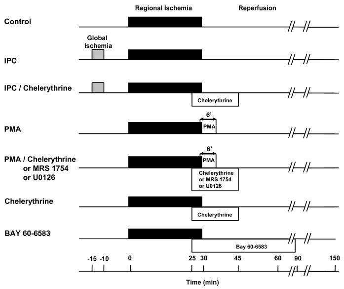
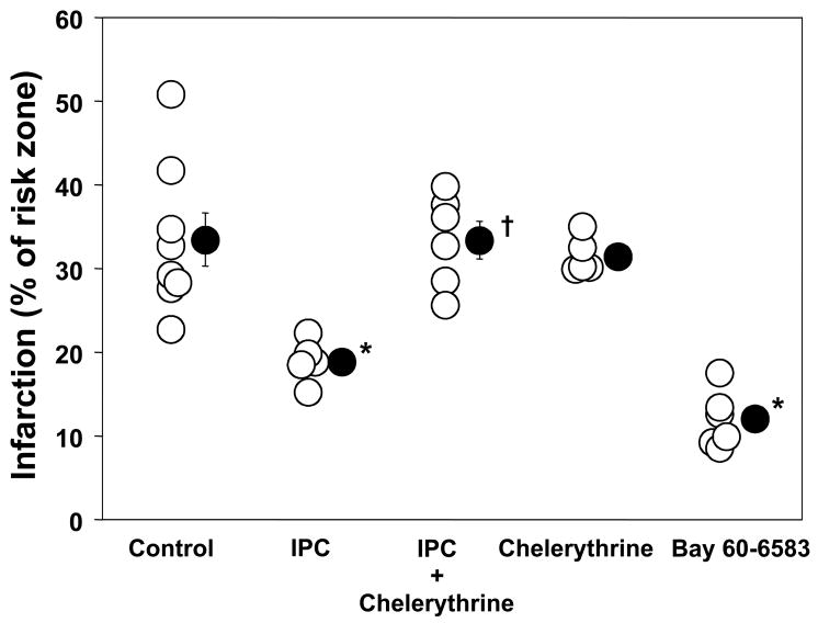
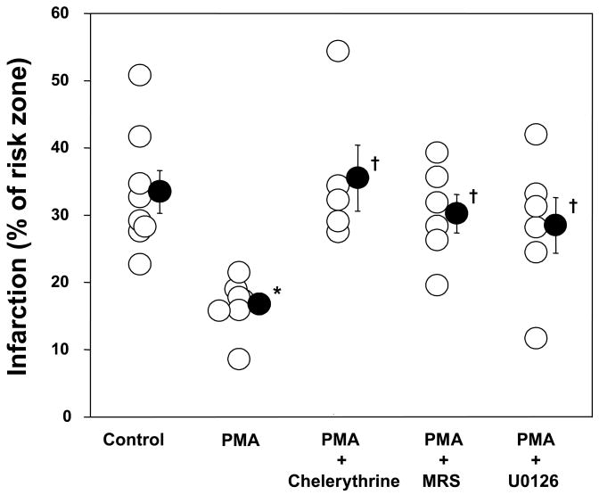
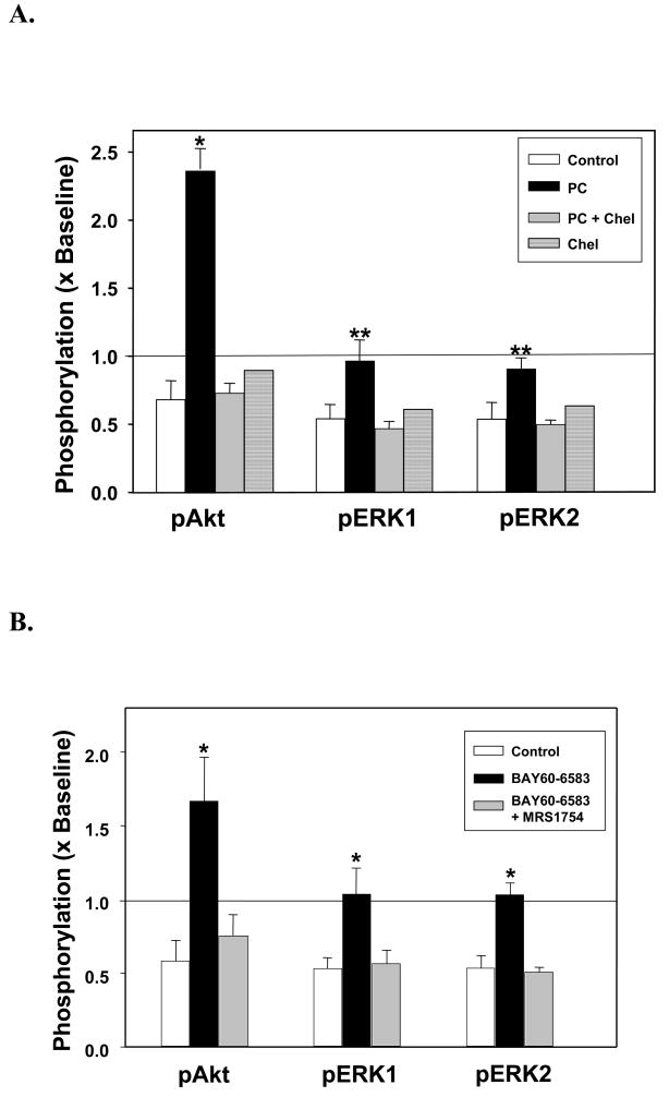
Although protein kinase C (PKC) plays a key role in ischemic preconditioning (IPC), the actual mechanism of that protection is unknown. We recently found that protection from IPC requires activation of adenosine receptors during early reperfusion. We, therefore, hypothesized that PKC might act to increase the heart's sensitivity to adenosine. IPC limited infarct size in isolated rabbit hearts subjected to 30-min regional ischemia/2-h reperfusion and IPC's protection was blocked by the PKC inhibitor chelerythrine given during early reperfusion revealing involvement of PKC at reperfusion. Similarly chelerythrine infused in the early reperfusion period blocked the increased phosphorylation of the protective kinases Akt and ERK1/2 observed after IPC. Infusing phorbol 12-myristate 13-acetate (PMA), a PKC activator, during early reperfusion mimicked IPC's protection. As expected, the protection triggered by PMA at reperfusion was blocked by chelerythrine, but surprisingly it was also blocked by MRS1754, an adenosine A(2b) receptor-selective antagonist, suggesting that PKC was somehow facilitating signaling from the A(2b) receptors. NECA [5'-(N-ethylcarboxamido) adenosine], a potent but not selective A(2b) receptor agonist, increased phosphorylation of Akt and ERK1/2 in a dose-dependent manner. Pretreating hearts with PMA or brief preconditioning ischemia had no effect on phosphorylation of Akt or ERK1/2 per se but markedly lowered the threshold for NECA to induce their phosphorylation. BAY 60-6583, a highly selective A(2b) agonist, also caused phosphorylation of ERK1/2 and Akt. MRS1754 prevented phosphorylation induced by BAY 60-6583. BAY 60-6583 limited infarct size when given to ischemic hearts at reperfusion. These results suggest that activation of cardiac A(2b) receptors at reperfusion is protective, but because of the very low affinity of the receptors endogenous cardiac adenosine is unable to trigger their signaling. We propose that the key protective event in IPC occurs when PKC increases the heart's sensitivity to adenosine so that endogenous adenosine can activate A(2b)-dependent signaling.


Figures

Figure 1
Figure 1
Infarct protocols. Abbreviations: IPC = ischemic preconditioning, PMA = phorbol 12-myristate 13-acetate
Figure 2
Figure 2
Biochemical protocols. Arrows indicate times of biopsies of left ventricle. Abbreviations: see Fig. 1, NECA = 5′-(N-ethylcarboxamido)adenosine, Chel = chelerythrine
Figure 3
Figure 3
Effect of ischemic preconditioning (IPC) and administration of a protein kinase C inhibitor chelerythrine during early reperfusion on myocardial infarct size as a percentage of the risk zone. BAY 60-6583 an A2b-selective adenosine agonist given at reperfusion also reduced infarct size. Open circles represent individual experiments while closed circles depict group mean±SEM. IPC’s anti-infarct effect was blocked by chelerythrine (2.8 μM). *p<0.05 vs. control, †p<0.05 vs. IPC
Figure 4
Figure 4
Effect of phorbol 12-myristate 13-acetate (PMA, 0.05 nM) and inhibitors on myocardial infarct size as a percentage of the risk zone. Open circles represent individual experiments while closed circles depict group mean±SEM. PMA at reperfusion reduced infarct size. A protein kinase C inhibitor chelerythrine, an adenosine A2b receptor-selective antagonist MRS 1754 (20 nM), and a MEK and therefore ERK1/2 inhibitor U0126 (0.5 μM) abolished PMA’s protection. *p<0.05 vs. control, †p<0.05 vs. PMA
Figure 5
Figure 5
Summary data of changes in phosphorylation of Akt (Ser473) and ERK1/2 (Thr202/Thr204) in hearts following 30 min of global ischemia and 10 min of reperfusion. Phosphorylation is normalized to that in a preischemic sample. (A) Phosphorylation of Akt and ERK in Control hearts (n=4) and hearts preconditioned (PC) with 5 min of global ischemia preceding the 30-min index ischemia with (n=4) or without (n=4) chelerythrine (Chel, 2.8 μM) during the 10 min of reperfusion. Measurements were also made in a non-preconditioned heart treated only with chelerythrine during reperfusion. *p<0.001 and **p<0.05 vs. Control and PC + Chel. (B) Phosphorylation of Akt and ERK in hearts after 10 min of reperfusion in Control hearts, hearts treated with BAY 60-6583 alone during reperfusion or hearts treated with BAY 60-6583 + MRS 1754. *p<0.05 vs. Control and BAY 60-6583+ MRS 1754.
Figure 6
Figure 6
Graph of Akt phosphorylation (Ser473) as a % of phospho-Akt at baseline after 5 min of either 2.5, 5.0 or 10 nM NECA infusion in escalating doses in 4 isolated hearts. A representative western blot of one of these hearts is seen at the top of the figure. Phospho-Akt significantly increased after infusion of 5.0 and 10 nM NECA, whereas 2.5 nM NECA did not change phosphorylation status of Akt. The A2a-selective agonist CGS 21680 at a concentration 3 times its Kd had no effect on Akt phosphorylation. *p<0.05 vs. baseline
Figure 7
Figure 7
(A) Representative western blots of bands for phospho-Akt (Ser473) in control hearts, hearts treated with phorbol 12-myristate 13-acetate (PMA, 0.05 nM) alone, PMA + MRS 1754, or hearts exposed to brief preconditioning ischemia. The left-hand column presents phospho-Akt bands before infusion of the subthreshold dose of NECA, while the right-hand column presents bands after NECA treatment. Phosphorylation increased after NECA administration only in hearts pre-treated with either PMA or IPC. MRS 1754 blocked any increase. (B) Phospho-Akt (Ser473) and (C and D) phospho-ERK1/2 (Thr202/Thr204) as % of levels measured just before treatment with NECA. All hearts in panels B–D were treated for 5 min with the subthreshold dose of 2.5 nM NECA. NECA had no effect in untreated hearts. After pretreatment with PMA the subthreshold dose of NECA now caused a robust phosphorylation of Akt and ERK1/2. NECA had no effect on phosphorylation when co-infused with a selective A2b receptor antagonist MRS 1754 in PMA treated hearts. Brief ischemia (IPC) also enabled the subthreshold dose of NECA to phosphorylate Akt and ERK1/2. Each bar is mean±SEM of 4 observations. *p<0.05 vs. control, #p<0.05 vs. PMA
Figure 8
Figure 8
(A) Effect of 300nM BAY 60-6583 on Akt and ERK phosphorylation in serial biopsies from isolated rabbit hearts taken at baseline, after 5 and 10 min of drug administration, and again after 10 min of washout. BAY 60-6583 nearly doubled phosphorylation of each. (B) Phosphorylation of Akt and ERK1/2 was blocked by MRS 1754. *p<0.05, †p<0.01, ‡p<0.005, all vs Baseline

Similar articles

Cited by

References

    1. Hausenloy DJ, Tsang A, Mocanu MM, Yellon DM. Ischemic preconditioning protects by activating prosurvival kinases at reperfusion. Am J Physiol. 2005;288:H971–H976. - PubMed
    1. Philipp S, Yang X-M, Cui L, Davis AM, Downey JM, Cohen MV. Postconditioning protects rabbit hearts through a protein kinase C-adenosine A2b receptor cascade. Cardiovasc Res. 2006;70:308–314. - PubMed
    1. Yang X-M, Proctor JB, Cui L, Krieg T, Downey JM, Cohen MV. Multiple, brief coronary occlusions during early reperfusion protect rabbit hearts by targeting cell signaling pathways. J Am Coll Cardiol. 2004;44:1103–1110. - PubMed
    1. Solenkova NV, Solodushko V, Cohen MV, Downey JM. Endogenous adenosine protects preconditioned heart during early minutes of reperfusion by activating Akt. Am J Physiol. 2006;290:H441–H449. - PubMed
    1. Kin H, Zatta AJ, Lofye MT, Amerson BS, Halkos ME, Kerendi F, et al. Postconditioning reduces infarct size via adenosine receptor activation by endogenous adenosine. Cardiovasc Res. 2005;67:124–133. - PubMed

Publication types

MeSH terms