

Repeat microsphere delivery for serial measurement of regional blood perfusion in the chronically instrumented, conscious canine
- PMID: 17632127
- PMCID: PMC2277484
- DOI: 10.1016/j.jss.2007.04.012
Repeat microsphere delivery for serial measurement of regional blood perfusion in the chronically instrumented, conscious canine
Abstract
Introduction: For chronic, repeat hemodynamic studies in conscious dogs, we designed and tested a chronically instrumented canine microsphere delivery model. The goals of this study were (1) to investigate the accuracy of repeated estimations of blood perfusion using fluorescent-labeled microspheres and (2) to develop and validate a chronic preparation that permits consecutive estimations in the same conscious animal over an extended protocol.
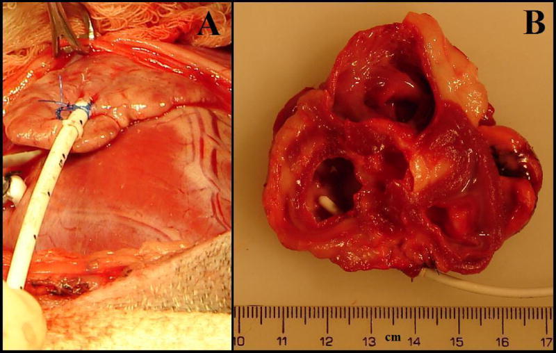
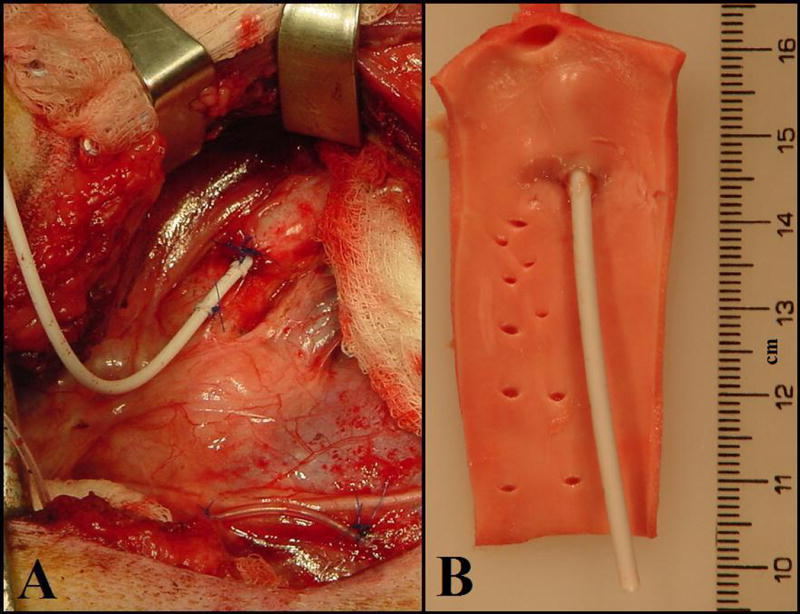
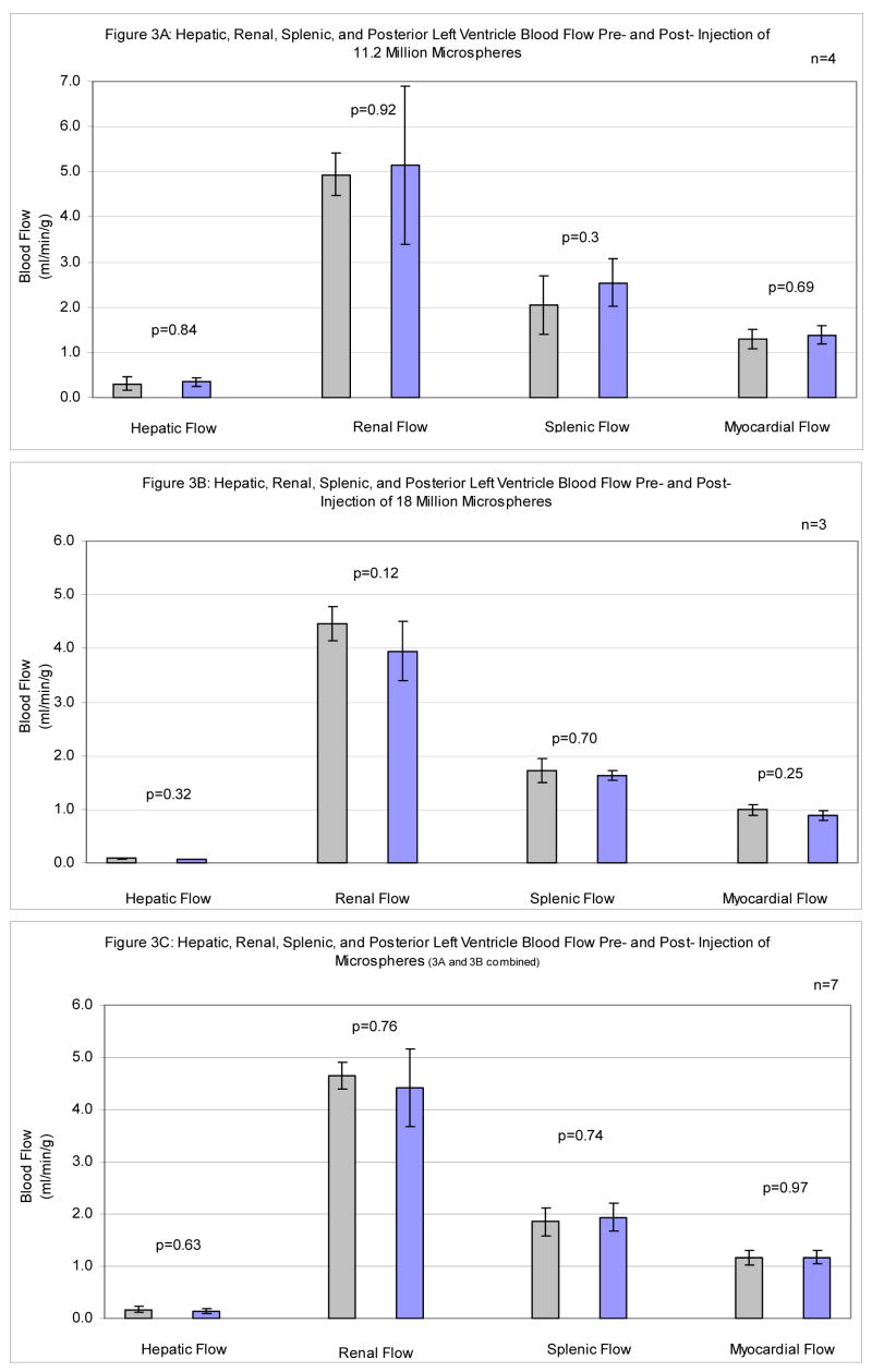
Methods: Via thoracotomy, nine dogs were instrumented with left atrial appendage and aortic vascular access catheters connected to subcutaneous vascular access ports. Four animals received seven serial injections of 1.6 million 15 microm microspheres (total: 11.2 million), and five animals received 8 serial injections of 2.25 million microspheres (total: 18 million) over the course of 11 or 18 wk.
Results: All catheters have remained bidirectionally patent during protocol for 14.9 +/- 0.8 (mean +/- SEM) wk. Sphere accumulation did not significantly alter global myocardial (P = 0.69, P = 0.25), renal (P = 0.92, P = 0.12), hepatic (P = 0.84, P = 0.32), or splenic (P = 0.33, P = 0.70) blood perfusion in either set of animals.
Conclusions: Catheters remained bidirectionally patent for months, did not interfere with the hemodynamic responses of the preparation, and allowed repeat percutaneous injection of microspheres and withdrawal of reference arterial blood from within conscious canines. Eight serial injections totaling 18 million microspheres over 18 weeks did not alter regional myocardial, hepatic, renal, or splenic blood flow. This dependable, chronic, percutaneous arterial access preparation provides a means for examining acute and long-term effects of pathophysiological, pharmaceutical, and environmental influences on regional arterial blood flow in conscious, large animals.
Figures
References
-
- Rudolph AM, Heymann MA. The circulation of the fetus in utero. Methods for studying distribution of blood flow, cardiac output and organ blood flow. Circ Res. 1967 Aug;21(2):163–84. - PubMed
-
- Makowski EL, Meschia G, Droegemueller W, Battaglia FC. Measurement of umbilical arterial blood flow to the sheep placenta and fetus in utero. Distribution to cotyledons and the intercotyledonary chorion. Circ Res. 1968 Nov;23(5):623–31. - PubMed
-
- Hale SL, Alker KJ, Kloner RA. Evaluation of nonradioactive, colored microspheres for measurement of regional myocardial blood flow in dogs. Circulation. 1988 Aug;78(2):428–34. - PubMed
-
- Glenny RW, Bernard S, Brinkley M. Validation of fluorescent-labeled microspheres for measurement of regional organ perfusion - PubMed
-
- Prinzen FW, Glenny RW. Developments in non-radioactive microsphere techniques for blood flow measurement. Cardiovasc Res. 1994 Oct;28(10):1467–75. - PubMed
Publication types
MeSH terms
Grants and funding
LinkOut - more resources
Full Text Sources
