. 2007 Nov 10;368(1):73-82.
doi: 10.1016/j.virol.2007.06.017. Epub 2007 Jul 16.

Replacement of the respiratory syncytial virus nonstructural proteins NS1 and NS2 by the V protein of parainfluenza virus 5

Affiliations

Replacement of the respiratory syncytial virus nonstructural proteins NS1 and NS2 by the V protein of parainfluenza virus 5

Kim C Tran et al. Virology. .

Abstract

Paramyxoviruses have been shown to produce proteins that inhibit interferon production and signaling. For human respiratory syncytial virus (RSV), the nonstructural NS1 and NS2 proteins have been shown to have interferon antagonist activity through an unknown mechanism. To understand further the functions of NS1 and NS2, we generated recombinant RSV in which both NS1 and NS2 were replaced by the PIV5 V protein, which has well-characterized IFN antagonist activities (DeltaNS1/2-V). Expression of V was able to partially inhibit IFN responses in DeltaNS1/2-V-infected cells. In addition, the replication kinetics of DeltaNS1/2-V were intermediate between DeltaNS1/2 and wild-type (rA2) in A549 cells. However, expression of V did not affect the ability of DeltaNS1/2-V to activate IRF3 nuclear translocation and IFNbeta transcription. These data indicate that V was able to replace some of the IFN inhibitory functions of the RSV NS1 and NS2 proteins, but also that NS1 and NS2 have functions in viral replication beyond IFN antagonism.


Figures

Figure 1
Figure 1
Generation of V mutant rRSV. (a) Genomic structure of V mutant rRSV. (b) Expression of viral proteins in rRSV-infected cells. Vero cells were infected by the indicated viruses at a MOI of 3. Cell lysates were harvested at 16 h p.i. and separated by SDS-PAGE. Viral proteins were detected by Western blot analysis using antibodies directed against RSV structural proteins (top), V (middle), or NS1 and NS2 (bottom). (c) Electropherograms of sequences of V editing sites. Total RNA was extracted from cells infected by PIV5, ΔNS1/2-Vwt, and ΔNS1/2-Vmut at a MOI of 3 for 16 hours and subjected to RT-PCR using the V cloning primers. PCR fragments were isolated and sequenced.
Figure 2
Figure 2
V expressed by rRSV causes STAT1 degradation. A549 cells were infected by rA2 (a), ΔNS1/2 (b), ΔNS1/2-Vwt (c), or ΔNS1/2-Vmut (d) at a MOI of 3. Total cell extracts were harvested at 4 hour intervals as indicated and subjected to Western blot analysis using antibodies to V and STAT1. N expression is shown as a control for infection.
Figure 3
Figure 3
Accumulation of IFNβ and ISG15 mRNA in rRSV-infected cells. A549 cells were infected with the indicated viruses at a MOI of 3. At 8, 12, and 16 h p.i., total RNA was isolated from the infected cells and subjected to Northern blot analysis using radiolabled DNA probes to IFNβ (top) or ISG15 (bottom). Bands were visualized by autoradiography. After exposure, the blots were stripped and reprobed with a radiolabeled probe for GAPDH as a loading control (lower panels).
Figure 4
Figure 4
Activation of IRF-3 in rRSV-infected cells. A549 cells on coverslips were infected at a MOI of 3 by the indicated viruses. At 9 (a), 12 (b), or 16 (c) h p.i., coverslips were fixed in formalin, permeabilized, and subjected to indirect immunofluorescence using antibodies to F (green) and IRF-3 (red). The percentages of infected cell displaying nuclear IRF-3 (graphs) were determined by counting approximately 300 cells per coverslip. Each sample was assayed in triplicate. Error bars represent SEM. (d) A549 cells were infected at a MOI of 3 by the indicated viruses. Total cell lysates were harvested 16 h p.i. and subjected to Western blot analysis for phosphorylated (Ser396) and total IRF3. (e) EMSA for IRF3 binding. A549 cells were infected at a MOI of 3 by the indicated viruses. Nuclear extracts were harvested at 16 h p.i. and used in an EMSA with a radiolabeled ISRE probe. CC, 100X cold competitor.
Figure 4
Figure 4
Activation of IRF-3 in rRSV-infected cells. A549 cells on coverslips were infected at a MOI of 3 by the indicated viruses. At 9 (a), 12 (b), or 16 (c) h p.i., coverslips were fixed in formalin, permeabilized, and subjected to indirect immunofluorescence using antibodies to F (green) and IRF-3 (red). The percentages of infected cell displaying nuclear IRF-3 (graphs) were determined by counting approximately 300 cells per coverslip. Each sample was assayed in triplicate. Error bars represent SEM. (d) A549 cells were infected at a MOI of 3 by the indicated viruses. Total cell lysates were harvested 16 h p.i. and subjected to Western blot analysis for phosphorylated (Ser396) and total IRF3. (e) EMSA for IRF3 binding. A549 cells were infected at a MOI of 3 by the indicated viruses. Nuclear extracts were harvested at 16 h p.i. and used in an EMSA with a radiolabeled ISRE probe. CC, 100X cold competitor.
Figure 5
Figure 5
Multiple step growth analysis of V mutant rRSV. A549 (a), Vero (b), HEp-2 (c), or HeLa (d) cells were infected with the V mutant rRSV at a MOI of 0.01 and supernatant samples were harvested daily for 5 d p.i. Supernatants were clarified and viral titers were determined. The analysis was performed in triplicate. Shown are the mean titers; error bars represent the SEM.
Figure 6
Figure 6
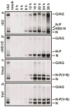
Viral RNA production in rRSV-infected cells. HEp-2 cells were infected by rA2 (a), ΔNS1/2 (b), ΔNS1/2-Vwt (c), or ΔNS1/2-Vmut (d) at a MOI of 3. Total cellular RNA was harvested at 4 hour intervals as indicated and subjected to Northern blot analysis using a radiolabeled DNA probe to RSV N. Viral RNA species are indicated on the right. G/AG, genome/antigenome.

Similar articles

Cited by

References

    1. Andrejeva J, Childs KS, Young DF, Carlos TS, Stock N, Goodbourn S, Randall RE. The V proteins of paramyxoviruses bind the IFN-inducible RNA helicase, mda-5, and inhibit its activation of the IFN-beta promoter. Proc Natl Acad Sci U S A. 2004;101(49):17264–9. - PMC - PubMed
    1. Andrejeva J, Poole E, Young DF, Goodbourn S, Randall RE. The p127 subunit (DDB1) of the UV-DNA damage repair binding protein is essential for the targeted degradation of STAT1 by the V protein of the paramyxovirus simian virus 5. J Virol. 2002a;76(22):11379–86. - PMC - PubMed
    1. Andrejeva J, Young DF, Goodbourn S, Randall RE. Degradation of STAT1 and STAT2 by the V proteins of simian virus 5 and human parainfluenza virus type 2, respectively: consequences for virus replication in the presence of alpha/beta and gamma interferons. J Virol. 2002b;76(5):2159–67. - PMC - PubMed
    1. Atreya PL, Peeples ME, Collins PL. The NS1 protein of human respiratory syncytial virus is a potent inhibitor of minigenome transcription and RNA replication. J Virol. 1998;72(2):1452–1461. - PMC - PubMed
    1. Beeler JA, van Wyke Coelingh K. Neutralization epitopes of the F glycoprotein of respiratory syncytial virus: effect of mutation upon fusion function. Journal of Virology. 1989;63(7):2941–2950. - PMC - PubMed

Publication types

MeSH terms

Substances