Review
. 2007 Aug 6;18(12):1207-13.
doi: 10.1097/WNR.0b013e328244e6c3.

Spatial remapping of the visual world across saccades

Affiliations
Review

Spatial remapping of the visual world across saccades

Paul M Bays et al. Neuroreport. .

Abstract

Recent research has identified neurons in the visual system that remap their receptive fields before a saccade. The activity of these neurons may signal a prediction of postsaccadic visual input, derived from an efference copy of saccadic motor output. Such a prediction is often thought to underlie our perception of a stable visual world, by compensating for the shifts in retinal image that accompany each eye movement. Here we review the evidence, and conclude that prediction does not in fact play a significant role in maintaining visual stability. Instead, we consider a novel perspective in which the primary function of spatial remapping is to support three key nonperceptual processes: action control, sensorimotor adaptation and spatial memory.


Figures

Figure 1
Figure 1
Detecting intrasaccadic changes to visual input. An eye movement (indicated by the white arrow, top) causes a global shift in the image falling on the retina. According to the spatial remapping model, an internal retinotopic representation of the visual scene (bottom) is translated by a distance and direction specified by an efference copy of the saccadic motor command. This remapping (indicated by the black arrows) results in a prediction of the expected visual input following the saccade. Comparison with the actual post-saccadic input reveals any discrepancy due to external motion (for instance, in this scene, movement of the two walkers). Where there is no discrepancy, the visual scene is perceived as stable.
Figure 2
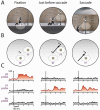
Figure 2
Neural basis of spatial remapping. A, An example visual neuron has a receptive field located just below fixation (left). Immediately before a saccade is made from ‘a’ to ‘b’ the neuron’s receptive field shifts to a new position (centre). This position corresponds to the expected location of the receptive field following the eye movement (right). B, Remapping is probed experimentally in monkeys by flashing a probe in either the pre-saccadic receptive field (RF) or the post-saccadic receptive field (future field, FF), at various times relative to the saccade. C, Activity of an example neuron in the frontal eye field. Firing rate (in spikes s-1) is aligned with probe onset, for probes presented at the times illustrated in A and B above. The visual response shifts from the RF to the FF just before the saccade. (Adapted with permission from [5].)
Figure 3
Figure 3
The double-step saccade task. Two targets (1 & 2) are briefly presented in quick succession. The subject must then saccade from the initial fixation point (F) to the remembered target locations in the correct sequence (solid arrows). Motor planning of the second saccade must take into account the change in eye position due to the first saccade. Planning the second eye movement without remapping the retinotopic representation of target 2 would result in an erroneous horizontally-deviated saccade (dashed arrow) computed on the basis of the second target’s retinotopic position as viewed originally at fixation point (F).
Figure 4
Figure 4
A, A typical experimental procedure demonstrating saccadic suppression of displacement. A saccade target (X) is presented on a blank screen at a location peripheral to fixation (circle). Triggered by onset of the saccade, the target shifts to a new location. Subjects are generally very poor at detecting the intrasaccadic displacement. B, In a variant of this procedure, the saccade target is briefly blanked at saccade onset, reappearing in its new location only after the saccade is complete. This manipulation greatly improves subjects’ ability to detect the displacement [28]. C, Saccades are variable in size and direction due to noise in motor output. Black dots illustrate typical distributions of actual saccadic endpoints about the intended endpoint (arrow tip), for saccades of 10° and 20° (arrows not to scale). Grey ellipses indicate areas expected to contain 95% of endpoints. Uncertainty over saccadic endpoint is greater in the direction parallel to the saccade than perpendicular to it, and scales with saccade size.
Figure 5
Figure 5
Typical scanpath of a patient with a right parietal lesion searching for target letter Ts amongst distractors. Note both the neglect of the left side of the search array and the many refixations of stimuli on the right. (Adapted with permission from [44].)

References

    1. von Holst E, Mittelstaedt H. Das Reafferenzprincip. Naturwissenschaft. 1950;37:464–476.
    1. Sperry RW. Neural basis of the spontaneous optokinetic response produced by visual inversion. J Comp Physiol Psychol. 1950;32:482–489. - PubMed
    1. Duhamel JR, Colby CL, Goldberg ME. The updating of the representation of visual space in parietal cortex by intended eye movements. Science. 1992;255(5040):90–92. - PubMed
    1. Colby CL, Goldberg ME. Space and attention in parietal cortex. Annu Rev Neurosci. 1999;22:319–349. - PubMed
    1. Sommer MA, Wurtz RH. Influence of the thalamus on spatial visual processing in frontal cortex. Nature. 2006;444(7117):374–377. - PubMed

Publication types