. 2007 Aug;3(8):480-5.
doi: 10.1038/nchembio.2007.9. Epub 2007 Jul 15.

Unusual transformations in the biosynthesis of the antibiotic phosphinothricin tripeptide

Affiliations

Unusual transformations in the biosynthesis of the antibiotic phosphinothricin tripeptide

Joshua A V Blodgett et al. Nat Chem Biol. 2007 Aug.

Abstract

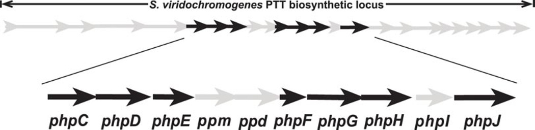
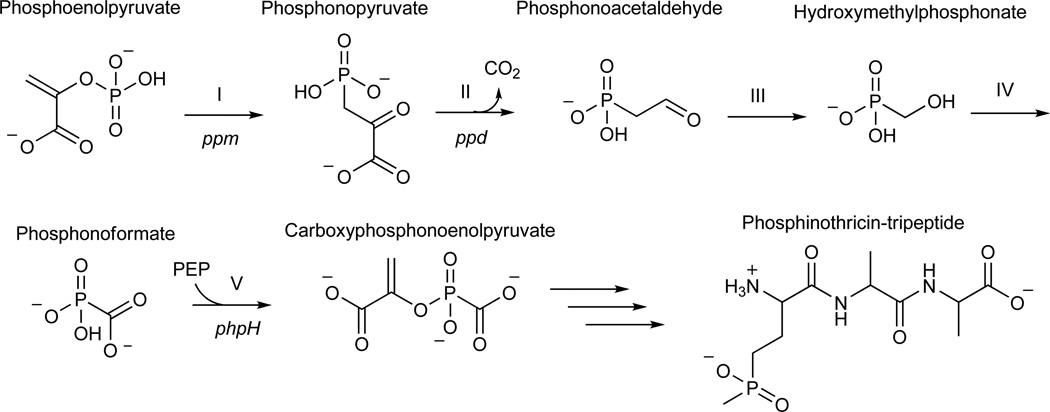
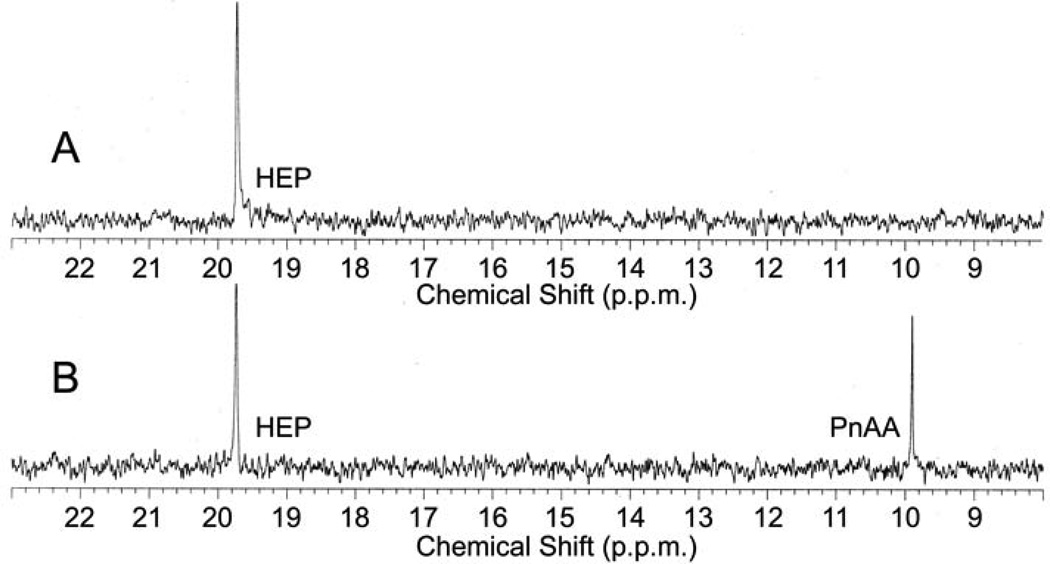
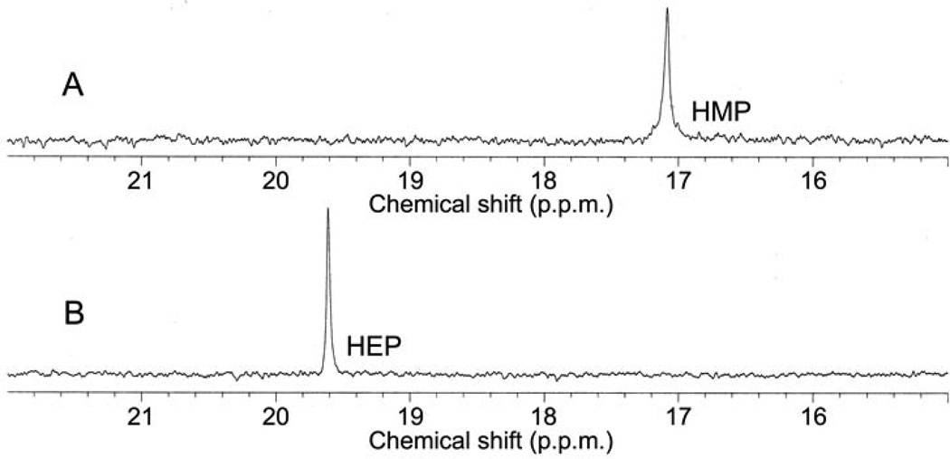
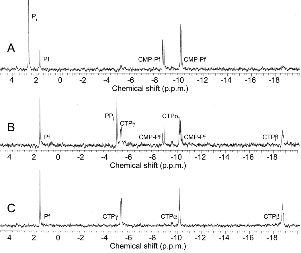
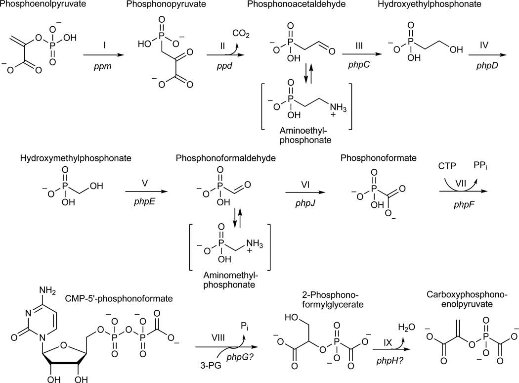
Phosphinothricin tripeptide (PTT, phosphinothricylalanylalanine) is a natural-product antibiotic and potent herbicide that is produced by Streptomyces hygroscopicus ATCC 21705 (ref. 1) and Streptomyces viridochromogenes DSM 40736 (ref. 2). PTT has attracted widespread interest because of its commercial applications and unique phosphinic acid functional group. Despite intensive study since its discovery in 1972 (see ref. 3 for a comprehensive review), a number of steps early in the PTT biosynthetic pathway remain uncharacterized. Here we report a series of interdisciplinary experiments involving the construction of defined S. viridochromogenes mutants, chemical characterization of accumulated intermediates, and in vitro assay of selected enzymes to examine these critical steps in PTT biosynthesis. Our results indicate that early PTT biosynthesis involves a series of catalytic steps that to our knowledge has not been described so far, including a highly unusual reaction for carbon bond cleavage. In sum, we define a pathway for early PTT biosynthesis that is more complex than previously appreciated.


Figures

Figure 1
Figure 1
Map of PTT biosynthetic genes and model for early steps in PTT biosynthesis. (a) Detail of genes in the core of the PTT biosynthetic gene cluster. Genes shown in grey were not studied in this work because they have experimentally established roles in PTT biosynthesis or because homology has allowed roles to be assigned by inference. The open reading frames in black did not have experimentally assigned roles, except for phpH, where this gene is expected to function in CPEP synthesis. (b) Model for carboxyphosphonoenolpyruvate biosynthesis from phosphonoacetaldehyde via the intermediates hydroxymethylphosphonate and phosphonoformate (adapted from). Steps referred to in the text are denoted by roman numerals, and S. viridochromogenes genes thought to be involved in each step are indicated where possible.
Figure 1
Figure 1
Map of PTT biosynthetic genes and model for early steps in PTT biosynthesis. (a) Detail of genes in the core of the PTT biosynthetic gene cluster. Genes shown in grey were not studied in this work because they have experimentally established roles in PTT biosynthesis or because homology has allowed roles to be assigned by inference. The open reading frames in black did not have experimentally assigned roles, except for phpH, where this gene is expected to function in CPEP synthesis. (b) Model for carboxyphosphonoenolpyruvate biosynthesis from phosphonoacetaldehyde via the intermediates hydroxymethylphosphonate and phosphonoformate (adapted from). Steps referred to in the text are denoted by roman numerals, and S. viridochromogenes genes thought to be involved in each step are indicated where possible.
Figure 2
Figure 2
In vitro reconstitution of early PTT biosynthetic reactions monitored by 31P NMR. (a) 31P NMR results of the His-PhpC catalyzed reduction of phosphonoacetaldehyde (PnAA, δ 9.9) to hydroxyethylphosphonate (HEP, δ 9.7) in the presence of (A) NADH and (B), the same reaction performed in the presence of NADPH. (b) 31P NMR results confirming the direct in vitro conversion of hydroxyethylphosphonate to hydroxymethylphosphonate by PhpD. (A), an assay performed in the presence of Rosetta(DE3)pLysS/pJVD26 extract containing PhpD, showing the conversion of hydroxyethylphosphonate into hydroxymethylphosphonate (HMP, δ 17.1). (B) a control assay containing hydroxyethylphosphonate (HEP, δ 19.6) and Rosetta(DE3)pLysS extract. (c) 31P NMR results of assays establishing the CTP-phosphonoformate nucleotidyltransferase activity of His-PhpF. (A) A reaction containing His-PhpF, inorganic pyrophosphatase, CTP and phosphonoformate showing the accumulation of phosphate (δ 2.6), and the conversion of substrates into CMP-5’-phosphonoformate (δ −8.6, −10.1). The top of the phosphate peak is not shown to fit the spectrum to the figure. (B), the same reaction as in A in the absence of pyrophosphatase showing the partial conversion of phosphonoformate and CTP into CMP-5’-phosphonoformate (δ −8.6, −10.1) and pyrophosphate (δ −4.7) and (C) a control assay containing heat-inactivated His-PhpF, CTP (δ −5.1, −9.97, −18.6) and phosphonoformate (δ 1.7), The putative α, β, and γ designations of the CTP phosphorus nuclei have been assigned by comparison to those analogously found in ATP .
Figure 2
Figure 2
In vitro reconstitution of early PTT biosynthetic reactions monitored by 31P NMR. (a) 31P NMR results of the His-PhpC catalyzed reduction of phosphonoacetaldehyde (PnAA, δ 9.9) to hydroxyethylphosphonate (HEP, δ 9.7) in the presence of (A) NADH and (B), the same reaction performed in the presence of NADPH. (b) 31P NMR results confirming the direct in vitro conversion of hydroxyethylphosphonate to hydroxymethylphosphonate by PhpD. (A), an assay performed in the presence of Rosetta(DE3)pLysS/pJVD26 extract containing PhpD, showing the conversion of hydroxyethylphosphonate into hydroxymethylphosphonate (HMP, δ 17.1). (B) a control assay containing hydroxyethylphosphonate (HEP, δ 19.6) and Rosetta(DE3)pLysS extract. (c) 31P NMR results of assays establishing the CTP-phosphonoformate nucleotidyltransferase activity of His-PhpF. (A) A reaction containing His-PhpF, inorganic pyrophosphatase, CTP and phosphonoformate showing the accumulation of phosphate (δ 2.6), and the conversion of substrates into CMP-5’-phosphonoformate (δ −8.6, −10.1). The top of the phosphate peak is not shown to fit the spectrum to the figure. (B), the same reaction as in A in the absence of pyrophosphatase showing the partial conversion of phosphonoformate and CTP into CMP-5’-phosphonoformate (δ −8.6, −10.1) and pyrophosphate (δ −4.7) and (C) a control assay containing heat-inactivated His-PhpF, CTP (δ −5.1, −9.97, −18.6) and phosphonoformate (δ 1.7), The putative α, β, and γ designations of the CTP phosphorus nuclei have been assigned by comparison to those analogously found in ATP .
Figure 3
Figure 3
An alternative pathway for carboxyphosphonoenolpyruvate biosynthesis from phosphonoacetaldehyde supported by data presented in this work. Steps referred to in the text are indicated by roman numerals and are annotated with their respective genetic determinants. Bracketed intermediates were identified from blocked mutants and likely represent modifications of the corresponding aldehyde intermediates.

References

    1. Seto H, et al. Studies on the biosynthesis of bialaphos (SF-1293). 1. Incorporation of 13C- and 2H-labeled precursors into bialaphos. The Journal of antibiotics. 1982;35(12):1719–1721. - PubMed
    1. Bayer E, et al. Phosphinothricin und Phosphinothricyl-Alanyl-Alanin. Helvetica Chimica Acta. 1972;55:224–239. - PubMed
    1. Thompson CJ, Seto H. In: Genetics and Biochemistry of Antibiotic Production. Vining LC, Stuttard C, editors. Newton, MA: Butterworth- Heinemann; 1995. pp. 197–222.
    1. Abell LM, Villafranca JJ. Investigation of the mechanism of phosphinothricin inactivation of Escherichia coli glutamine synthetase using rapid quench kinetic technique. Biochemistry. 1991;30:6135–6141. - PubMed
    1. Bailey RR. Brief overview of single-dose therapy for uncomplicated urinary tract infections. Chemotherapy. 1990;36(Suppl 1):27–30. - PubMed

Publication types

MeSH terms