. 2007 Aug;14(8):696-703.
doi: 10.1038/nsmb1272. Epub 2007 Jul 15.

The positions of TFIIF and TFIIE in the RNA polymerase II transcription preinitiation complex

Affiliations

The positions of TFIIF and TFIIE in the RNA polymerase II transcription preinitiation complex

Hung-Ta Chen et al. Nat Struct Mol Biol. 2007 Aug.

Abstract

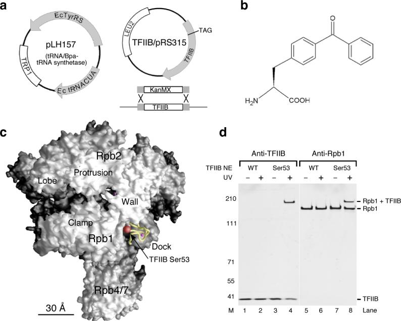
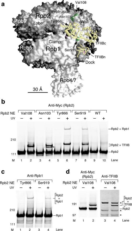
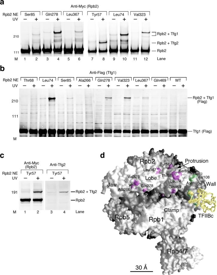
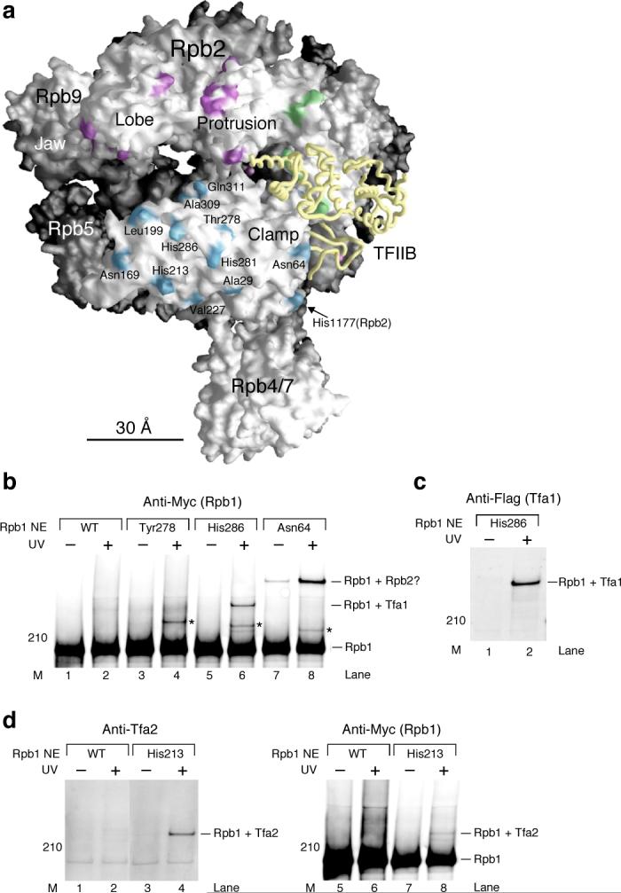
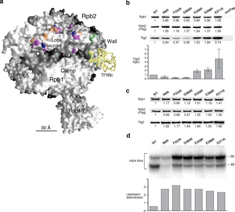
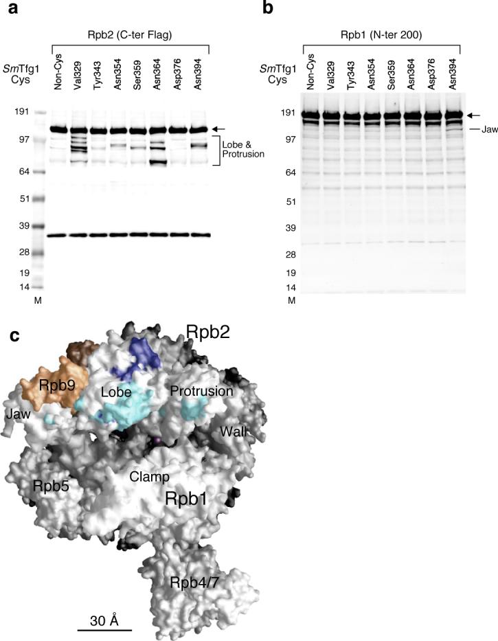
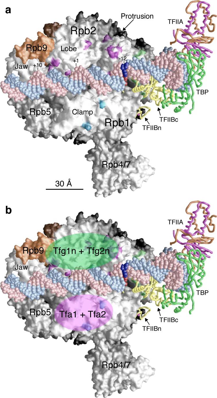
We incorporated the non-natural photoreactive amino acid p-benzoyl-L-phenylalanine (Bpa) into the RNA polymerase II (Pol II) surface surrounding the central cleft formed by the Rpb1 and Rpb2 subunits. Photo-cross-linking of preinitiation complexes (PICs) with these Pol II derivatives and hydroxyl-radical cleavage assays revealed that the TFIIF dimerization domain interacts with the Rpb2 lobe and protrusion domains adjacent to Rpb9, while TFIIE cross-links to the Rpb1 clamp domain on the opposite side of the Pol II central cleft. Mutations in the Rpb2 lobe and protrusion domains alter both Pol II-TFIIF binding and the transcription start site, a phenotype associated with mutations in TFIIF, Rpb9 and TFIIB. Together with previous biochemical and structural studies, these findings illuminate the structural organization of the PIC and the network of protein-protein interactions involved in transcription start site selection.


Figures

Figure 1
Figure 1. Incorporation of the non-natural amino acid p-Benzoyl-L-Phenylalanine (Bpa) to TFIIB and photocrosslinking
(a) Two plasmids in the TFIIB amber strain. Plasmid pLH157 (Trp1+) contains orthogonal tRNA synthetase (Ec TyrRS) and tRNA (Ec tRNACUA) genes for incorporating Bpa through nonsense suppression of the TAG codon. Plasmid TFIIB/pRS315 (Leu2+) is transformed through plasmid shuffling into the yeast strain containing replacement of chromosomal TFIIB by the KanMX marker. (b) The chemical structure of the photocrosslinker Bpa. (c) Position of the TFIIB ribbon domain on the Pol II surface. The TFIIB ribbon (yellow backbone with a magenta sphere representing the zinc atom) is modeled onto the surface of a 12-subunit Pol II structure based on superposition of the Pol II-IIB co-crystal structure (PDB code: 1R5U10) with two other Pol II structures: a 12-subunit Pol II (PDB code: 1WCM18) and a Pol II elongation complex (PDB code: 1SFO42). This model of the Pol II surface is used throughout the figures. Location of Ser53 on the ribbon domain is shown with the red sphere representing the entire serine residue including backbone and side-chain atoms. All structural figures were generated using Grasp. (d) Western analysis of TFIIB-Bpa crosslinking within the PIC. As indicated, TFIIB Ser53-Bpa is crosslinked to Rpb1. The fusion protein band (Rpb1+TFIIB) is recognized by the two antibodies (see METHODS). TFIIB NE: TFIIB-Bpa nuclear extract. UV: UV irradiation. M: molecular weight marker.
Figure 2
Figure 2. Photocrosslinking confirms the binding between the TFIIB core domain and the Rpb2 wall domain within the PIC
(a) Molecular surface of Pol II and model of TFIIB position within the PIC. The TFIIB core domain (TFIIBc, yellow backbone) is modeled on the Pol II surface based on previous site-specific biochemical analyses using IIB cysteine mutants. The TFIIB ribbon is modeled as in Fig 1c. The locations of Bpa on the Rpb2 wall are shown with the green colored patches on the surface. The magenta sphere within the central cleft of Pol II denotes the Mg atom inside the active site. (b) Western analysis of Rpb2 photocrosslinking. Rpb2 and crosslinking bands were revealed with anti-Myc antibody against the Myc epitope-tagged Rpb2. As indicated, the crosslinked proteins were identified as TFIIB and Rpb1 (see below, (c) and (d)). Rpb2 NE: Rpb2-Bpa nuclear extract. UV: UV irradiation. M: molecular weight marker. Asterisk: unidentified crosslinking target. (c) Identification of the crosslinked protein Rpb1. Anti-Rpb1 antibody against the first 200 amino acids of Rpb1 was used to validate the crosslinking target for Tyr866- and Ser919-Bpa. (d) Representative validation of Rpb2-TFIIB crosslinking. The antibody against TFIIB was used to reveal TFIIB in the crosslinking band (Rpb2+TFIIB) from Rpb2 Val108-Bpa. Asterisk: non-TFIIB background.
Figure 3
Figure 3. TFIIF binds the Rpb2 lobe and protrusion domains within the PIC
(a) Western analysis of Rpb2 photocrosslinking. Anti-Myc antibody against the Myc epitope tagged Rpb2 was used to reveal Rpb2 and crosslinking products. As indicated, Tfg1 and Tfg2 subunits of TFIIF were identified as the crosslinked proteins (see below, (b) and (c)). Rpb2 NE: Rpb2-Bpa nuclear extract. M: molecular weight marker. UV: UV irradiation. (b) Tfg1 was identified as the crosslinked polypeptide with Rpb2-Bpa mutants. Identification was based the method using the recombinant TFIIF containing Flag epitope tagged Tfg1 (see METHODS). Flag tagged Tfg1 was revealed by Western analysis using the anti-Flag antibody. The crosslinking bands (Rpb2+Tfg1) were also shown to contain Myc epitope tagged Rpb2 by staining with the anti-Myc antibody (data not shown). (c) Tfg2 crosslinked to Bpa positioned at Tyr57 of Rpb2. Anti-Myc antibody was used to reveal Myc epitope tagged Rpb2 and crosslinking bands, and anti-Tfg2 antibody was used to validate the crosslinked Tfg2. (d) Locations of Rpb2-Bpa crosslinked to TFIIF. Purple patches on the Pol II surface indicate Rpb2-TFIIF crosslinking positions. Same as Figure 2a, Bpa positions crosslinked with TFIIB are colored green and the TFIIB core domain (TFIIBc) is shown as the yellow backbone. The magenta sphere within the central cleft of Pol II denotes the magnesium atom in the active site.
Figure 4
Figure 4. TFIIE interacts with the Rpb1 clamp domain within the PIC
(a) Positions of Bpa in the Rpb1 clamp domain are displayed as the light blue patches on the Pol II surface. One Bpa position (His1177) belongs to the Rpb2 subunit. Positions of Rpb2-Bpa crosslinked to TFIIF (Fig. 3; Tfg1 and Tfg2) and TFIIB (Fig. 2) are colored purple and green, respectively. TFIIB backbone is colored yellow. The magenta sphere inside the central cleft denotes the active site Mg atom. (b) Western analysis of photocrosslinking using Rpb1-Bpa mutants. Rpb1 and crosslinking bands were revealed by the anti-Myc antibody against Myc epitope tagged Rpb1. As indicated, the crosslinked protein for His286-Bpa was identified as Tfa1 (see below, (c)). One of the crosslinked proteins for Asn64-Bpa is likely Rpb2 (Rpb1+Rpb2?) based on the structure of Pol II. Asterisk: crosslinked protein has not been identified. Rpb1 NE: Rpb1-Bpa nuclear extract. M: molecular weight marker. UV: UV irradiation. (c) The crosslinked protein for His286-Bpa is identified to be Tfa1. Identification of Rpb1-Tfa1 crosslinking is based on the mixing method described in METHODS. Anti-Flag antibody was used to reveal Tfa1. The same crosslinking band was also recognized by the anti-Myc antibody against Myc epitope tagged Rpb1 (data not shown). (d) The crosslinked protein for His213-Bpa is Tfa2. Anti-Tfa2 antibody and anti-Myc antibody (Rpb1) were used in the Western analysis to validate Rpb1-Tfa2 crosslinking.
Figure 5
Figure 5. Rpb2 lobe/protrusion mutations alter Pol II-IIF interaction and confer an upstream shift in the transcription start site
(a) Model of the Pol II surface and mutations in the Rpb2 lobe and protrusion domains. Mutations of Rpb2 that demonstrated slow growth phenotype are colored orange. Lethal mutations are colored blue. Purple and green patches denote Bpa positions that are crosslinked to TFIIF (Fig. 3) and TFIIB (Fig. 2), respectively. TFIIB backbone (TFIIBc; TFIIB core domain) is colored yellow. (b) Mutations in the Rpb2 lobe and protrusion domains affect TFIIF binding. Co-immune precipitation was conducted with the anti-Flag agarose gel to pull down TFIIF and Pol II containing Flag epitope tagged Rpb2. Rpb2 mutations are indicated on top. Anti-Rpb1, anti-Flag (Rpb2) and anti-Tfg2 antibodies were used in the Western analysis. The relative intensity of immune staining was normalized against the wild-type (WT) and is listed below each protein band. NonFlag: Wild-type Rpb2 without Flag epitope tag. The intensity ratio between Tfg2 and Rpb1 is displayed in the bottom panel. Error bars indicate s.e.m. (n=3). (c) PIC formation is not affected by the Rpb2 mutations. The immobilized template assay (PIC formation) using the Rpb2 mutant nuclear extracts was analyzed by Western blotting. The relative intensity of each protein was normalized as described in (b). (d) Pol II transcription initiation is affected by the Rpb2 lobe and protrusion mutants. The transcription start sites for the Rpb2 mutants were analyzed by in vitro transcription and primer extension. All Rpb2 mutants shift the preference for transcription start to the upstream site (−60; relative to the ATG translation start codon of the HIS4 gene). The ratio of upstream and downstream start sites (−60/−49) is shown in the lower panel.
Figure 6
Figure 6. Site-specific hydroxyl radical protein cleavage reveals the binding site for the TFIIF dimerization domain within the PIC
(a) Cleavage fragments of Rpb2 with the Flag epitope attached at the C-terminus were visualized by Western analysis with anti-Flag antibody. Cysteine mutation sites in SmTfg1 recombinant TFIIF cys variants are indicated. A non-Cys SmTfg1 is included as a control. The specific cleavage fragments correspond to cut sites in the Rpb2 lobe and protrusion domains. The arrow points to full length Rpb2. M; molecular size marker. (b) Cleavage fragments of Rpb1 were visualized by Western analysis with antibody against the first 200 residues of Rpb1. SmTfg1 FeBABE derivatives are indicated. The specific cleavage fragment in N394C corresponds to cut sites in the Rpb1 jaw domain. The arrow points to full length Rpb1. (c) The calculated FeBABE cleavage sites (Supp Figs 4,5) for SmTfg1 dimerization domain Cys variants were mapped to the surface of the Pol II. A nine-residue segment centered at the calculated cleavage site is colored light blue (weak cleavage) or dark blue (strong/medium cleavage). The magenta sphere located deep inside the active site represents the active site Mg. Rpb9 is colored orange.
Figure 7
Figure 7. Model of PIC assembly
(a) Model of the complex is based on TFIIB core domain (TFIIBc) binding to Pol II from previous biochemical analyses and non-natural amino acid photocrosslinking in this study. TFIIA, TBP, and DNA were fitted into the complex based on the crystal structures of human TFIIB core/TBP/TATA box ternary complex (PDB code: 1C9B32) and yeast TFIIA/TBP/TATA box complex (PDB code: 1RM135). TFIIB ribbon domain is modeled onto the Pol II surface based on the Pol II-TFIIB co crystal (PDB code: 1R5U), and the magenta sphere represents the zinc atom. TFIIB and TBP are shown as yellow and green backbone models, respectively. The two subunits of TFIIA are colored orange (Toa1) and magenta (Toa2). The DNA strands flanking the TATA box (recognized by TBP) is extended from the human TFIIBc/TBP/DNA x-ray structure using the straight B-form DNA. The DNA non-template strand is colored pink and the template strand is colored light blue. DNA base pair −12 (numbering based on the Adenovirus Major Late promoter used in the crystal structure), the site of DNA strand melting for Open complex formation, is colored dark red for the template strand and dark blue for the non-template strand, respectively. The DNA extends from promoter position −47 to +18 using the numbering of the Adenovirus Major Late promoter where +1 base is the transcription start and the TATA box sequence starts at −31. The Pol II surface is colored white except for Rpb9 (orange). Bpa positions crosslinked to TFIIB are colored green. Bpa positions crosslinked to TFIIF and TFIIE are colored purple and light blue, respectively. (b) Structural model is the same as (a). The locations of TFIIF dimerization domain (Tfg1n+ Tfg2n) and TFIIE (Tfa1+Tfa2) are shown with semi-transparent light green and magenta ovals, respectively.

Comment in

Similar articles

Cited by

References

    1. Hahn S. Structure and mechanism of the RNA polymerase II transcription machinery. Nat Struct Mol Biol. 2004;11:394–403. - PMC - PubMed
    1. Giardina C, Lis JT. DNA melting on yeast RNA polymerase II promoters. Science. 1993;261:759–62. - PubMed
    1. Pinto I, Wu WH, Na JG, Hampsey M. Characterization of sua7 mutations defines a domain of TFIIB involved in transcription start site selection in yeast. J Biol Chem. 1994;269:30569–73. - PubMed
    1. Sun ZW, Hampsey M. Identification of the gene (SSU71/TFG1) encoding the largest subunit of transcription factor TFIIF as a suppressor of a TFIIB mutation in Saccharomyces cerevisiae. Proc Natl Acad Sci U S A. 1995;92:3127–31. - PMC - PubMed
    1. Chen BS, Hampsey M. Functional interaction between TFIIB and the Rpb2 subunit of RNA polymerase II: implications for the mechanism of transcription initiation. Mol Cell Biol. 2004;24:3983–91. - PMC - PubMed

Publication types