

Review
doi: 10.1038/labinvest.3700656.
Epub 2007 Jul 16.
Diversity in cell surface sialic acid presentations: implications for biology and disease
Affiliations
- PMID: 17632542
- PMCID: PMC7100186
- DOI: 10.1038/labinvest.3700656
Item in Clipboard
Review
Diversity in cell surface sialic acid presentations: implications for biology and disease
Lab Invest.
2007 Sep.
Abstract
Sialic acids (Sias) are typically found as terminal monosaccharides attached to cell surface glycoconjugates. They play many important roles in many physiological and pathological processes, including microbe binding that leads to infections, regulation of the immune response, the progression and spread of human malignancies and in certain aspects of human evolution. This review will provide some examples of these diverse roles of Sias and briefly address immunohistochemical approaches to their detection.
Figures
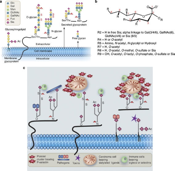
Natural diversity in Sia presentation, structure and function. (a) Diversity in presentation. Sias are typically found at the terminal position of N- and O-linked glycans attached to the cell surface and to secreted glycoproteins, as well as on glycosphingolipids expressed at the cell surface. Ac, O-acetyl ester; Fuc, fucose; Gal, galactose; GalNAc, N-acetylgalactosamine; Glc, glucose; GlcNAc, N-acetylglucosamine; Man, mannose; S, sulfate ester. Reproduced with permission from Varki. (b) Structural diversity. All Sias share the common feature of having nine carbons, a carboxylic acid residue at the 1-position, and a variety of linkages to the underlying sugar chain from the 2-position. Various types of substitutions at the 4, 5, 7, 8 and 9 positions combine with the linkage variation to generate the diversity of Sias found in nature. Only a portion of this diversity is represented here. Some examples of the binding specificity of probes for detection of Sia types and linkages are discussed in the Box 1. (c) Examples of pathobiological interactions involving Sias. This cartoon shows examples of the types of interactions involving recognition of Sia diversity. The molecules and cells are obviously not drawn to scale.
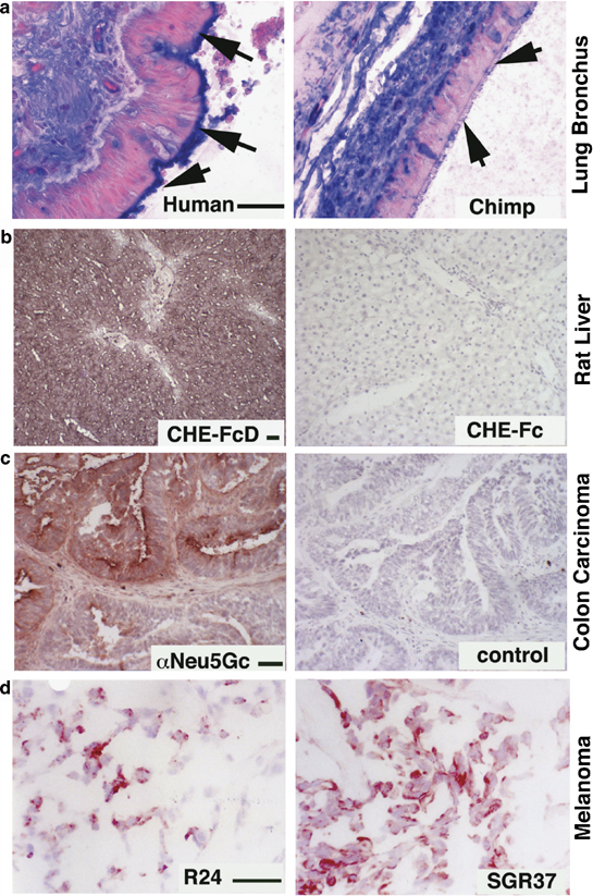
Some Examples of in situ detection of Sia types and linkages. (a) Differential display of α2-6-linked Sias on the ciliary border of lung bronchial epithelial cells in humans and chimpanzees. Paraffin sections were deparaffinized, rehydrated and blocked for endogenous biotin and overlaid with biotinylated Sambucus Nigra agglutinin (SNA), followed by Tris-buffered saline washes, alkaline phosphatase-labeled streptavidin and additional washes. Color detection used a Vector Labs blue substrate and nuclear fast red counterstain. Arrows point to the epithelial luminal edge, where staining is seen in humans, but not in chimpanzees or in other great apes. (b) Expression of 9-O-acetylated Sias in rat liver. Left panel: DFP-treated recombinant soluble influenza C Hemagglutinin-esterase (CHE-FcD). Right panel: non-DFP-treated version of the same probe (CHE-Fc, esterase active, negative binding control). Frozen sections of rat liver were blocked for endogenous peroxidases, fixed in formalin, washed, overlaid with the CHE-FcD or Che-Fc probes, which were pre-complexed with HRP anti-human antibodies at predetermined dilutions. After incubation at 4°C for 2 h, sections were washed in PBS and color developed using the AEC substrate with Mayer's hematoxylin for nuclear counterstain. (c) Detection of the nonhuman Sia Neu5Gc in human tumors, using an affinity-purified chicken IgY antibody. Left Panel: example of positive colon carcinoma. Frozen sections of a human colon carcinoma were blocked for endogenous peroxidases, fixed in formalin, washed and overlaid with a chicken anti-Neu5Gc antibody. Following washes and incubation with HRP-labeled donkey anti-chicken antibody and further washes, color was developed using the AEC substrate with Mayer's hematoxylin for nuclear counterstain. The right panel shows a negative control using a nonspecific chicken IgY. (d) Detection of GD3 and de-N-acetylated GD3 in human melanomas. Left Panel: detection of GD3 using MAb R24. Right Panel: detection of de-N-acetyl-GD3 using MAb SGR37. Unfixed frozen sections of a human melanoma were blocked for endogenous peroxidases and overlaid with monoclonal antibodies R24 or SGR37. Following PBS washes, incubation with HRP-labeled goat anti-mouse Ig antibodies and further washes, color was developed using the AEC substrate with Mayer's hematoxylin for nuclear counterstain. (Note: Scale bar=50 μm for each pair of panels is shown on the left side).
References
-
- Varki A, Cummings R, Esko JD. Essentials of Glycobiology. Cold Spring Harbor Laboratory Press: Plainview, NY; 1999. - PubMed
-
- Drickamer K, Taylor M. Introduction to Glycobiology. Oxford University Press: Oxford, UK; 2006.
Publication types
MeSH terms
Substances
LinkOut - more resources
Full Text Sources
Other Literature Sources
