Review
. 2007 Aug;5(8):598-610.
doi: 10.1038/nrmicro1704.

Viral RNA pseudoknots: versatile motifs in gene expression and replication

Affiliations
Review

Viral RNA pseudoknots: versatile motifs in gene expression and replication

Ian Brierley et al. Nat Rev Microbiol. 2007 Aug.

Abstract

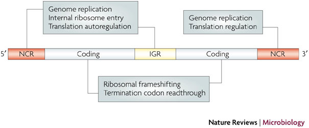
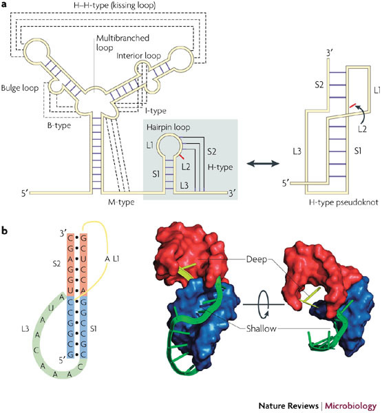
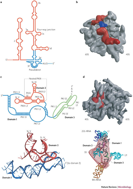
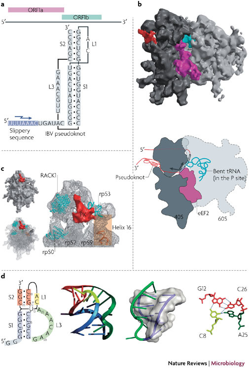
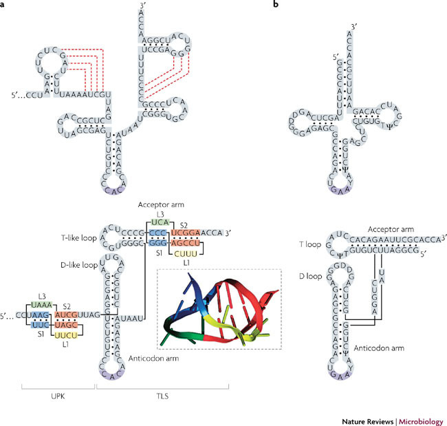
RNA pseudoknots are structural elements found in almost all classes of RNA. First recognized in the genomes of plant viruses, they are now established as a widespread motif with diverse functions in various biological processes. This Review focuses on viral pseudoknots and their role in virus gene expression and genome replication. Although emphasis is placed on those well defined pseudoknots that are involved in unusual mechanisms of viral translational initiation and elongation, the broader roles of pseudoknots are also discussed, including comparisons with relevant cellular counterparts. The relationship between RNA pseudoknot structure and function is also addressed.


Conflict of interest statement

The authors declare no competing financial interests.

Figures

Figure 1
Figure 1. RNA pseudoknots in virus gene expression.
A schematic of a generic RNA virus genome is shown. Viral pseudoknots have been described in the 5′ non-coding region (NCR), the coding region, the intergenic region (IGR) and the 3′ NCR, where they function in various steps of the replication cycle. Although the majority of examples are from positive-strand RNA viruses, pseudoknots also have a role in the replication cycles of certain DNA viruses, satellite RNA viruses and viroids. For simplicity, viral pseudoknots involved in long-range interactions (including virus genome circularization) or possessing catalytic activity are not shown, but are discussed in the text.
Figure 2
Figure 2. RNA pseudoknot structure.
a | Various structural motifs have been described in RNA. Orthodox secondary structures consist of base-paired regions (stems) connected by single-stranded loops at stem termini (hairpin loop), or in the body of a stem (bulge (B) or interior (I) loop) or at the junction of several stems (multibranched (M) loop). Pseudoknots are considered as a tertiary structure and form when bases in a loop pair with a single-stranded region elsewhere. The hairpin type (H-type) pseudoknot is by far the most common, and this tertiary interaction involves bases in the loop of a hairpin loop. The resultant structure contains two stem regions, S1 and S2, connected by single-stranded loops. In many cases, no unpaired bases are present between the two stems (L2 is zero), and the stems stack coaxially to give a quasi-continuous helix. b | The secondary structure of the pseudoknot of the ribosomal frameshifting signal of simian retrovirus 1 (SRV-1) is shown alongside three dimensional views of the nuclear magnetic resonance model. The stems are shown as surface representations and the loops as ribbons (all structural images were prepared using PyMol). The polarity and handedness of the double helix leads to inequivalence of the loops, with L1 (yellow) crossing the deep groove and L3 (green) crossing the shallow groove. S1 is blue, S2 is red, L1 is yellow and L3 is green. L2 is not present in the example shown.
Figure 3
Figure 3. Pseudoknots and internal ribosome entry.
a | A secondary structure representation of the hepatitis C virus (HCV) internal ribosome entry site (IRES) with the pseudoknot shown in blue. b | A surface representation of the human 80S ribosome (grey) in complex with the HCV IRES (red) derived from the cryo-electron microscopy (cryo-EM) structure. Density corresponding to the pseudoknot is indicated in blue. c | A secondary structure representation of the Plautia stali intestine virus (PSIV) IRES is shown above a ribbon representation of the RNA (domains 1 and 2) derived from the crystal structure. Domain 3 remains to be solved. The secondary structure of the cricket paralysis virus (CrPV) IRES (not shown) is similar. d | A surface representation of the yeast 80S ribosome (grey) is shown in complex with the CrPV IRES (red) derived from the cryo-EM structure. Below is a fit of the density to the modelled CrPV IRES showing the interactions that occur between the various domains and ribosomal components. Ribosomal proteins are cyan, 25S ribosomal RNA (rRNA) is purple and 18S rRNA is brown.
Figure 4
Figure 4. Pseudoknots and ribosomal frameshifting.
a | The overlapping coding sequences open reading frame 1a (ORF1a) and ORF1b of the genome of the coronavirus infectious bronchitis virus (IBV) are shown above the minimal frameshift-promoting sequences of this virus. The pseudoknot promotes frameshifting at the slippery sequence, indicated by a jagged arrow. b | A representation of the stalled, pseudoknot-engaged rabbit 80S ribosome is shown derived from the cryo-electron microscopy structure. The 60S subunit is light grey and the 40S subunit is dark grey. The peptidyl (P)-site transfer RNA (tRNA) stalled in the complex is coloured turquoise, the eukaryotic translocase, elongation factor 2 (eEF2), is purple and the pseudoknot structure is red. Below is a schematic of the stalled ribosome. Engagement with the pseudoknot generates a frameshifting intermediate in which the ribosome is stalled during translocation with eEF2 bound, generating tension in the mRNA that bends the P-site tRNA in a(+) sense direction. As a result, the anticodon–codon interaction breaks over the slippery sequence, allowing a spring-like relaxation of the tRNA in a (−) sense direction. c | A close-up view of the pseudoknot (PK) in the stalled complex, with the ribosomal components (rpS0, rpS2, rpS3 and rpS9) in close proximity highlighted. d | The beet western yellows virus (BWYV) pseudoknot is illustrated to show examples of features that might confound the ribosomal helicase. Shown from left to right are: a secondary structure model of the pseudoknot, with U13 drawn to indicate its extrusion form the helix; a ribbon representation of the X-ray structure; the L3–S1 (loop 3–stem 1) triplex interaction, with S1 shown as a transparent surface and the helix within as a purple ribbon; and the BWYV junction quadruple interaction, with hydrogen bonds shown as dashed lines. A triplex also forms at the junction of the two stems (not highlighted). RACK1, receptor for activated C kinase 1. Panels b and c are modified with permission from Ref. © MacMillan Publishers Ltd.
Figure 5
Figure 5. Pseudoknots and transfer RNA-like structures.
a | The 3′ end of the turnip yellow mosaic virus (TYMV) genomic RNA. In the upper panel, the predicted secondary structure is shown, with pseudoknotting interactions indicated by dashed red lines. Below is a secondary structure representation of the folded molecule, showing the transfer RNA (tRNA)-like structure (TLS) and the pseudoknots in the acceptor arm and upstream of the TLS (UPK). The ribbon representation (boxed) is derived from the nuclear magnetic resonance structure of the acceptor arm pseudoknot. b | For comparison, secondary structure representations of tRNAPhe are also shown. D, dihydrouridine modified bases; T, ribothymidine base.

Similar articles

Cited by

References

    1. Rietveld K, Van Poelgeest R, Pleij CW, Van Boom JH, Bosch L. The tRNA-like structure at the 3′ terminus of turnip yellow mosaic virus RNA. Differences and similarities with canonical tRNA. Nucleic Acids Res. 1982;10:1929–1946. doi: 10.1093/nar/10.6.1929. - DOI - PMC - PubMed
    1. Rietveld K, Pleij CW, Bosch L. Three-dimensional models of the tRNA-like 3′ termini of some plant viral RNAs. EMBO J. 1983;2:1079–1085. doi: 10.1002/j.1460-2075.1983.tb01549.x. - DOI - PMC - PubMed
    1. Pleij CW, Rietveld K, Bosch L. A new principle of RNA folding based on pseudoknotting. Nucleic Acids Res. 1985;13:1717–1731. doi: 10.1093/nar/13.5.1717. - DOI - PMC - PubMed
    1. Pleij CW. Pseudoknots: a new motif in the RNA game. Trends Biochem. Sci. 1990;15:143–147. doi: 10.1016/0968-0004(90)90214-V. - DOI - PubMed
    1. ten Dam E, Pleij K, Draper D. Structural and functional aspects of RNA pseudoknots. Biochemistry. 1992;31:11665–11676. doi: 10.1021/bi00162a001. - DOI - PubMed