

Brain image registration using cortically constrained harmonic mappings
- PMID: 17633713
- PMCID: PMC4511383
- DOI: 10.1007/978-3-540-73273-0_30
Brain image registration using cortically constrained harmonic mappings
Abstract
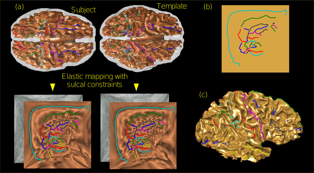
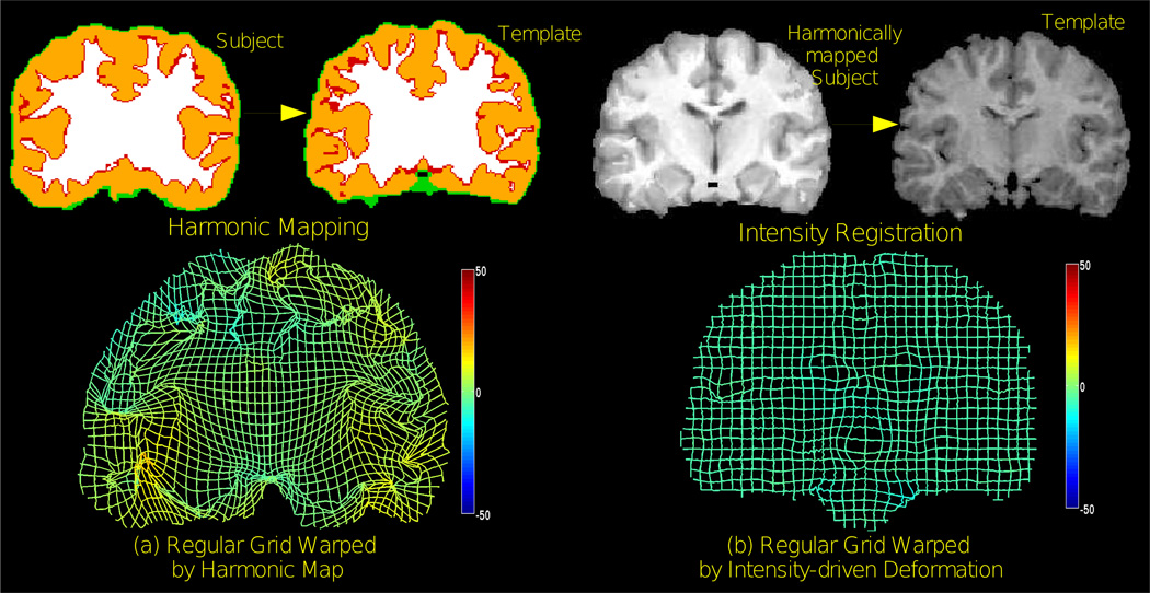
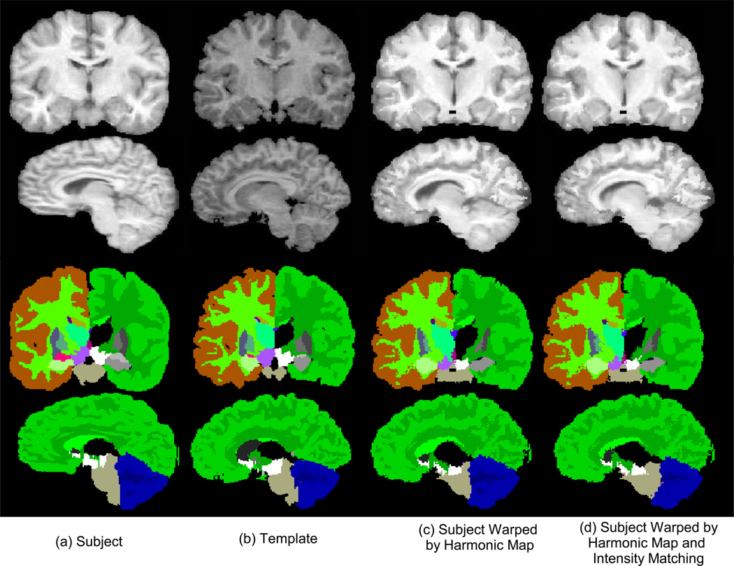
Volumetric registration of brains is required for inter-subject studies of functional and anatomical data. Intensity-driven registration typically results in some degree of misalignment of cortical and gyral folds. Increased statistical power in group studies may be achieved through improved alignment of cortical areas by using sulcal landmarks. In this paper we describe a new volumetric registration method in which cortical surfaces and sulcal landmarks are accurately aligned. We first compute a one-to-one map between the two cortical surfaces constrained by a set of user identified sulcal curves. We then extrapolate this mapping from the cortical surface to the entire brain volume using a harmonic mapping procedure. Finally, this volumetric mapping is refined using an intensity driven linear elastic registration. The resulting maps retain the one-to-one correspondence between cortical surfaces while also aligning volumetric features via the intensity-driven registration. We evaluate performance of this method in comparison to other volumetric registration methods.
Figures
Similar articles
-
A framework for brain registration via simultaneous surface and volume flow.Inf Process Med Imaging. 2009;21:576-88. doi: 10.1007/978-3-642-02498-6_48. Inf Process Med Imaging. 2009. PMID: 19694295 Free PMC article.
-
Surface-constrained volumetric brain registration using harmonic mappings.IEEE Trans Med Imaging. 2007 Dec;26(12):1657-69. doi: 10.1109/tmi.2007.901432. IEEE Trans Med Imaging. 2007. PMID: 18092736 Free PMC article.
-
Geometry driven volumetric registration.Inf Process Med Imaging. 2007;20:675-86. doi: 10.1007/978-3-540-73273-0_56. Inf Process Med Imaging. 2007. PMID: 17633739
-
Brain functional localization: a survey of image registration techniques.IEEE Trans Med Imaging. 2007 Apr;26(4):427-51. doi: 10.1109/TMI.2007.892508. IEEE Trans Med Imaging. 2007. PMID: 17427731 Review.
-
A review of 3D/2D registration methods for image-guided interventions.Med Image Anal. 2012 Apr;16(3):642-61. doi: 10.1016/j.media.2010.03.005. Epub 2010 Apr 13. Med Image Anal. 2012. PMID: 20452269 Review.
Cited by
-
A framework for brain registration via simultaneous surface and volume flow.Inf Process Med Imaging. 2009;21:576-88. doi: 10.1007/978-3-642-02498-6_48. Inf Process Med Imaging. 2009. PMID: 19694295 Free PMC article.
-
Combined volumetric and surface registration.IEEE Trans Med Imaging. 2009 Apr;28(4):508-22. doi: 10.1109/TMI.2008.2004426. Epub 2008 Aug 15. IEEE Trans Med Imaging. 2009. PMID: 19273000 Free PMC article.
-
Surface-guided computing to analyze subcellular morphology and membrane-associated signals in 3D.ArXiv [Preprint]. 2023 Apr 12:arXiv:2304.06176v1. ArXiv. 2023. PMID: 37090235 Free PMC article. Preprint.
-
Surface-guided computing to analyze subcellular morphology and membrane-associated signals in 3D.bioRxiv [Preprint]. 2023 Apr 20:2023.04.12.536640. doi: 10.1101/2023.04.12.536640. bioRxiv. 2023. PMID: 37131779 Free PMC article. Preprint.
-
Cortical and subcortical gray matter changes in patients with chronic tinnitus sustaining after vestibular schwannoma surgery.Sci Rep. 2021 Apr 16;11(1):8411. doi: 10.1038/s41598-021-87915-3. Sci Rep. 2021. PMID: 33863965 Free PMC article.
References
-
- Talairach J, Tournoux P. Co-planar Stereotaxic Atlas of the Human Brain: 3-Dimensional Proportional System - an Approach to Cerebral Imaging. Thieme Medical Publishers; New York, NY: 1988.
-
- Ashburner J, Friston K. Spatial normalization. In: Toga A, editor. Brain Warping. Academic Press; 1999. pp. 27–44.
-
- Woods RP, Grafton ST, Holmes CJ, Cherry SR, Mazziotta JC. Automated image registration: I. General methods and intrasubject, intramodality validation. Journal of Computer Assisted Tomography. 1998;22:139–152. - PubMed
-
- Hill DLG, Batchelor PG, Holden M, Hawkes DJ. Medical image registration. Phys. Med. Biol. 2001 Mar;46(4):R1–R45. - PubMed
-
- Christensen GE, Rabbitt RD, Miller MI, Joshi SC, Grenander U, Coogan TA, Essen DCV. Topological properties of smooth anatomic maps. IPMI. 1995:101–112.
Publication types
MeSH terms
Grants and funding
LinkOut - more resources
Full Text Sources
Other Literature Sources
Medical