

Horizontal transmission of Marek's disease virus requires US2, the UL13 protein kinase, and gC
- PMID: 17634222
- PMCID: PMC2045466
- DOI: 10.1128/JVI.01065-07
Horizontal transmission of Marek's disease virus requires US2, the UL13 protein kinase, and gC
Abstract
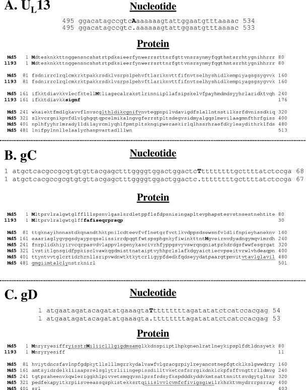
Marek's disease virus (MDV) causes a general malaise in chickens that is mostly characterized by the development of lymphoblastoid tumors in multiple organs. The use of bacterial artificial chromosomes (BACs) for cloning and manipulation of the MDV genome has facilitated characterization of specific genes and genomic regions. The development of most MDV BACs, including pRB-1B-5, derived from a very virulent MDV strain, involved replacement of the US2 gene with mini-F vector sequences. However, when reconstituted viruses based on pRB-1B were used in pathogenicity studies, it was discovered that contact chickens housed together with experimentally infected chickens did not contract Marek's disease (MD), indicating a lack of horizontal transmission. Staining of feather follicle epithelial cells in the skins of infected chickens showed that virus was present but was unable to be released and/or infect susceptible chickens. Restoration of US2 and removal of mini-F sequences within viral RB-1B did not alter this characteristic, although in vivo viremia levels were increased significantly. Sequence analyses of pRB-1B revealed that the UL13, UL44, and US6 genes encoding the UL13 serine/threonine protein kinase, glycoprotein C (gC), and gD, respectively, harbored frameshift mutations. These mutations were repaired individually, or in combination, using two-step Red mutagenesis. Reconstituted viruses were tested for replication, MD incidence, and their abilities to horizontally spread to contact chickens. The experiments clearly showed that US2, UL13, and gC in combination are essential for horizontal transmission of MDV and that none of the genes alone is able to restore this phenotype.
Figures


Similar articles
-
A full UL13 open reading frame in Marek's disease virus (MDV) is dispensable for tumor formation and feather follicle tropism and cannot restore horizontal virus transmission of rRB-1B in vivo.Vet Res. 2007 May-Jun;38(3):419-33. doi: 10.1051/vetres:2007009. Epub 2007 Mar 13. Vet Res. 2007. PMID: 17506972
-
Comparative sequence analysis of a highly oncogenic but horizontal spread-defective clone of Marek's disease virus.Virus Genes. 2007 Dec;35(3):753-66. doi: 10.1007/s11262-007-0157-1. Epub 2007 Aug 25. Virus Genes. 2007. PMID: 17721813
-
The Tegument Protein pUL47 of Marek's Disease Virus Is Necessary for Horizontal Transmission and Is Important for Expression of Glycoprotein gC.J Virol. 2020 Dec 22;95(2):e01645-20. doi: 10.1128/JVI.01645-20. Print 2020 Dec 22. J Virol. 2020. PMID: 32999032 Free PMC article.
-
Marek's disease virus research in the post-sequencing era: new tools for the study of gene functions and virus-host interactions.Avian Pathol. 2003 Aug;32(4):323-33. doi: 10.1080/0307945031000121068. Avian Pathol. 2003. PMID: 17585455 Review.
-
Marek's disease virus: lytic replication, oncogenesis and control.Expert Rev Vaccines. 2006 Dec;5(6):761-72. doi: 10.1586/14760584.5.6.761. Expert Rev Vaccines. 2006. PMID: 17184215 Review.
Cited by
-
Fluorescently tagged pUL47 of Marek's disease virus reveals differential tissue expression of the tegument protein in vivo.J Virol. 2012 Mar;86(5):2428-36. doi: 10.1128/JVI.06719-11. Epub 2011 Dec 21. J Virol. 2012. PMID: 22190714 Free PMC article.
-
Visualization of Marek's Disease Virus Genomes in Living Cells during Lytic Replication and Latency.Viruses. 2022 Jan 29;14(2):287. doi: 10.3390/v14020287. Viruses. 2022. PMID: 35215880 Free PMC article.
-
mRNA Splicing of UL44 and Secretion of Alphaherpesvirinae Glycoprotein C (gC) Is Conserved among the Mardiviruses.Viruses. 2024 May 15;16(5):782. doi: 10.3390/v16050782. Viruses. 2024. PMID: 38793663 Free PMC article.
-
Dual infection and superinfection inhibition of epithelial skin cells by two alphaherpesviruses co-occur in the natural host.PLoS One. 2012;7(5):e37428. doi: 10.1371/journal.pone.0037428. Epub 2012 May 21. PLoS One. 2012. PMID: 22629393 Free PMC article.
-
Purinergic signaling during Marek's disease in chickens.Sci Rep. 2023 Feb 4;13(1):2044. doi: 10.1038/s41598-023-29210-x. Sci Rep. 2023. PMID: 36739336 Free PMC article.
References
-
- Blondeau, C., N. Chbab, C. Beaumont, K. Courvoisier, N. Osterrieder, J. F. Vautherot, and C. Denesvre. 2007. A full UL13 open reading frame in Marek's disease virus (MDV) is dispensable for tumor formation and feather follicle tropism and cannot restore horizontal spread in rRB-1B in vivo. Vet. Res. 38:419-433. - PubMed
-
- Calnek, B. W. 2001. Pathogenesis of Marek's disease virus infection, p. 25-55. In K. Hirai (ed.), Marek's disease. Springer, Berlin, Germany. - PubMed
Publication types
MeSH terms
Substances
Grants and funding
LinkOut - more resources
Full Text Sources
Other Literature Sources
Miscellaneous
